
Nephrology
Abstract
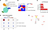
After encountering allogeneic nonself, monocytes differentiate to DCs, which in turn activate the adaptive immune system that drives transplant rejection. The downstream mechanisms of allorecognition are largely unknown. We analyzed scRNA-seq data sets to identify transcriptional changes in monocytes occurring after allostimulation. Hspa1a, which encodes HSP70, was upregulated in monocytes after allostimulation in contrast to syngeneic controls in mice lacking T, B, and NK cells. Similar findings were seen in scRNA-seq data derived from kidney biopsies of rejecting transplant patients. To validate the role of HSP70 in innate allorecognition and transplantation, we performed allogeneic bone marrow plug and kidney transplantation into WT, Hspa1a/Hspa1b–/–, and DSG-treated (HSP70 inhibitor) B6 mice and examined the graft immune infiltrate. Histology, T cell infiltration, and survival were assessed in kidney transplanted mice. In both models, DSG-treated or HSP70-KO recipients displayed significantly reduced infiltration by monocyte-derived DCs (mo-DC). Chronic rejection of Balb/c kidney grafts in WT B6 recipients was attenuated in DSG-treated and HSP70-KO recipients as indicated by a reduction in Banff score. Further experiments demonstrated that in vitro allostimulated immature HSP70-KO BMDC showed less maturation compared with WT BMDC. Targeting HSP70 in innate immune cells offers a novel approach to reduce chronic kidney graft rejection.
Authors
Zeping Gui, Neda Feizi, Berkay Demirors, Canxiang Lin, Hehua Dai, Mouhamad Al Moussawy, Andrew Friday, Steven M. Sanders, Latha Halesha, Amanda L. Williams, Faruk Sacirbegovic, Parmjeet S. Randhawa, Khodor I. Abou-Daya, Martin H. Oberbarnscheidt
Abstract
Missense mutations in the UMOD gene are a well-established genetic cause of autosomal dominant tubulointerstitial kidney disease (ADTKD). Here, we report two ADTKD pedigrees carrying de novo UMOD mutations, p.His36Tyr (H36Y) and p.Trp31Cys (W31C). To elucidate the underlying pathogenic mechanisms and explore targeted therapeutic strategies, we generated a UMODH36Y/+ knock-in mouse model using CRISPR/Cas9 technology, which recapitulated the major clinical manifestations observed in patients. Through single-cell RNA sequencing and experimental validation, we demonstrated that uromodulin mutations triggered endoplasmic reticulum (ER) stress and the unfolded protein response, with ferroptosis identified as the predominant mode of cell death in this disease. These findings were further confirmed in plasmid-transfected cell models expressing UMODH36Y and UMODW31C. Moreover, the molecular chaperone HSPA1A was identified as a potential therapeutic target. Geranylgeranylacetone, targeting HSPA1A as a chaperone drug, effectively mitigated endoplasmic reticulum stress, alleviated ferroptosis, and delayed the progression of renal dysfunction. Based on novel uromodulin mutations, our study reveals a pathogenic ER-ferroptosis axis in ADTKD-UMOD, deepens the insight into the pathogenesis of ADTKD-UMOD, and provides a promising strategy for developing chaperone therapy with drug repurposing in genetic kidney diseases.
Authors
Wen Shi, Yan Yang, Xianli Wen, Qianqian Wu, Jinxuan Wei, Xin-Lu Wang, Junyuan Shen, Siqi Peng, Xiaoliang Zhang, Bi-Cheng Liu, Bin Wang
Abstract
ADAMTS9 mutations cause the ciliopathies nephronophthisis and Joubert syndrome. Here we demonstrated that deletion of ADAMTS9 in the proximal nephron led to polycystic kidney development in mice. In males, Adamts9 deletion caused kidneys to become highly cystic while remaining small without undergoing enlargement. In contrast, female mice developed cystic kidneys at a slower rate. ADAMTS9 deletion disrupted ciliogenesis through the loss of cleavage of the ciliary transition zone (TZ) protein TMEM67, which led to loss of the MKS/B9 module – a key component of the ciliary gate. Functional analysis of all eight ciliopathy patient variants of ADAMTS9 identified to date showed TMEM67 C-terminus failed to localize to the TZ, thus disrupting a key regulatory mechanism in patient renal ciliogenesis. Modeling ADAMTS9-mediated TMEM67 cleavage utilizing TMEM67-cleavage deficient mice revealed loss of TZ formation, but not elevated canonical Wnt signaling as the underlying mechanism driving cystogenesis. Adamts9 deletion led to comparatively intense interstitial collagen deposition, which likely restricted kidney enlargement and resulted in the characteristically small kidney phenotype seen in nephronophthisis. By comparative analysis of four interconnected polycystic kidney models, in addition to Pkd1 and Pkd2 deleted kidneys, we identified differential collagen homeostasis as a principle factor determining cystic kidney size and type.
Authors
Sydney Fischer, Karyn L. Robert, Manu Ahmed, Griffin I. Kane, Matthew A. Kavanaugh, Wei Wang, Pamela V. Tran, Prabhani U. Atukorale, Sumeda Nandadasa

Abstract
Authors
Long Qian, Megan L. Baker, Laura Aponte Becerra, Cathleen Liang, Emma Koval, Kyra Shelton, Daris Javed, Gilbert Moeckel, Deepika Kumar, Avi Z. Rosenberg, Michael Kuperman, Tinyi Chu, Wassim Obeid, Xuefei Tian, Chirag R. Parikh, Leyuan Xu, Jonathan Barasch, Shuta Ishibe, Lloyd G. Cantley, Dennis G. Moledina
Abstract
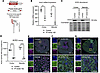
ACE2 is a membrane-bound monocarboxypeptidase strongly expressed in the renal proximal tubule (PT) with high affinity to degrade the vasopressor angiotensin II (AngII). We employed a mouse model of PT-specific ACE2 deletion (PT ACE2–KO) to demonstrate that the renal PT is a critical site for ACE2 regulation of blood pressure (BP) via modulation of the intrarenal renin-angiotensin system (RAS). While deletion of ACE2 from the PT had a minimal effect on baseline physiology, PT ACE2–KO mice were more susceptible to AngII hypertension than control mice. At day 5 of AngII infusion, the enhanced BP response was associated with cardiac hypertrophy, increased renal AngII levels, failure to suppress epithelial sodium channel (ENaC) γ cleavage, and increased sodium pump activity in PT ACE2–KO mice. Control mice instead increased renal ACE2 expression to reduce renal AngII accumulation and suppress intrarenal RAS activation, which offered protection from hypertension and complications. Transcriptional analysis corroborated changes in intrarenal RAS components and revealed alterations in distinct physiological pathways during AngII hypertension in PT ACE2–KO mice. Our studies provide evidence for alterations in ENaC regulation to contribute to the development of AngII hypertension and support PT-derived ACE2 as an integral member of the intrarenal RAS.
Authors
Jacqueline M. Emathinger, Zhidan Xiang, Jonathan W. Nelson, Matthew W. Hagen, Nakyung Kim, David I. Ortiz-Melo, Natalie Mattocks, Jorge F. Giani, Dexter L. Lee, Hannah Hartman-Houstman, Donna L. Ralph, Alicia A. McDonough, Brianna Chen, Stan G. Louie, Thomas M. Coffman, Susan B. Gurley
Abstract
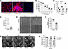
Autosomal dominant polycystic kidney disease (ADPKD) is a leading genetic cause of kidney failure, characterized by progressive cyst growth, inflammation, and interstitial fibrosis. Renal fibrosis, driven by myofibroblast activation and excessive extracellular matrix (ECM) deposition, is increasingly recognized as a key contributor to disease progression, yet targeted antifibrotic therapies remain limited. Here, we evaluated the therapeutic potential of pirfenidone to suppress fibrosis and disease progression in ADPKD. Single-nucleus RNA sequencing of human ADPKD kidneys identified fibroblasts as the predominant source of fibrous and adhesive ECM, with higher ECM-associated gene expression compared with that in normal kidney fibroblasts. In vitro, primary human ADPKD renal myofibroblasts displayed a similar profibrotic gene expression profile, and pirfenidone treatment suppressed ECM gene expression, cell proliferation, migration, and contractility. In the Pkd1RC/RC mouse model of ADPKD, pirfenidone reduced renal fibrosis, myofibroblast accumulation, ECM deposition, profibrotic gene expression, and associated signaling pathways and improved kidney function. Pirfenidone also reduced kidney enlargement but reduced cyst burden only in female mice. Collectively, these findings demonstrate that pirfenidone attenuates renal fibrosis and improves kidney function in ADPKD by suppressing myofibroblast activation and ECM production, supporting fibrosis as a therapeutic target and highlighting pirfenidone as a potential adjunct to cyst-directed therapies.
Authors
Viji Remadevi, Abeda Jamadar, Meekha M. Varghese, Haichun Yang, Sumedha Gunewardena, Darren P. Wallace, Reena Rao
Abstract
TGF-β is a central driver of kidney fibrosis, a common pathological hallmark of chronic kidney disease (CKD). Initiation of TGF-β signaling requires not only its synthesis but also the conversion of latent TGF-β to its bioactive form. However, the mechanisms governing TGF-β activation in the kidney and their contribution to kidney fibrosis remain poorly understood. Glycoprotein A repetitions predominant (GARP) anchors latent TGF-β on the cell surface and facilitates its bioactive release. Here, we show that GARP-mediated TGF‐β activation promotes kidney fibrosis. GARP was upregulated in both human and mouse CKD kidneys, predominantly in fibroblasts, and was induced by TNF in an NF-kB-dependent fashion. In multiple mouse models of kidney fibrosis, either global or fibroblast-specific deletion of GARP significantly reduced fibrosis. Mechanistically, GARP enables sustained production of active TGF-β, thereby amplifying fibroblast stimulation. Deletion of GARP in kidney fibroblasts lowered active TGF-β levels and attenuated fibroblast activation, whereas GARP overexpression enhanced TGF-β signaling. Notably, tamoxifen-induced deletion of GARP after fibrosis onset attenuated kidney fibrosis. Together, our findings identify GARP-mediated release of active TGF‐β as a critical step in sustaining fibroblast activation during kidney fibrosis and highlight GARP as a promising therapeutic target for CKD.
Authors
Yintong Chen, Weiwei Xu, Jieli Yu, Pei Deng, Nianping Liu, Yinyin Li, Hui Zhou, Hong Zhou, Jianchuan Wang, Bo Zhao, Florian Winau, Fan Fan Hou, Yu Hu
Abstract
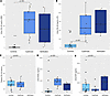
Early diagnosis of cystinosis is critical to limit disease progression. YKL-40, a protein in the chitinase family, released by inflammatory cells, may be a useful biomarker for cystinosis. In a case-control study of 10 children with cystinosis and 20 without cystinosis, matched by age and baseline eGFR, we measured urine YKL-40, NGAL, and EGF. A lateral flow device (LFD) for YKL-40 was also developed and tested. Urine YKL-40 was over 200-fold higher in children with cystinosis (64.6 ng/mL [IQR: 23.4, 83.8]) compared with controls (0.3 [IQR: 0.3, 0.79]; P = 0.0001) with excellent diagnostic discrimination (AUC = 0.99) that was superior to other biomarkers. LFD measurements for YKL-40 showed similar results (AUC = 0.93). YKL-40 results were verified in 5 cystinosis patients, and YKL-40 staining was markedly higher in kidney biopsies from cystinosis patients than in healthy controls. Urine YKL-40 has excellent diagnostic potential for cystinosis, and point-of-care technologies may facilitate early screening and management of this disease.
Authors
Jason H. Greenberg, Serena D Souza, Heather R. Thiessen Philbrook, Wassim Obeid, Avi Z. Rosenberg, Elena Levtchenko, Koenraad Veys, Susan L. Furth, Chirag R. Parikh
Abstract
Autoimmune kidney diseases can cause glomerulonephritis and tubulointerstitial nephritis, which if unresolved, lead to progressive glomerulosclerosis and tubulointerstitial fibrosis. The IL-1 receptor (IL-1R1) is known to have divergent and cell-specific effects in kidney injury. We hypothesized that IL-1R1 would dampen pro-inflammatory activation of myeloid cells such that deletion of myeloid cell IL-1R1 would exacerbate autoimmune nephritis. Mice with myeloid cell-specific deletion of IL-1R1 (LysMCre(+) / Il1r1fl/fl - MKO) and littermate controls (LysMCre(-) / Il1r1fl/fl - MWT) were subjected to nephrotoxic serum (NTS) nephritis. MKO mice demonstrated worsened glomerular and tubular injury as indicated by increased albuminuria, glomerular injury scores, and kidney mRNA levels of kidney injury molecule (KIM)-1 (Havcr1) and neutrophil gelatinase-associated lipocalin (NGAL/Lcn2). We further found that myeloid IL-1R1 deficiency resulted in increased myeloid cell ER stress and expression of the heterodimeric cytokine Ebi3/Il27a (IL-27). IL-27 then induced increased type I IFN expression by kidney endothelial cells. In turn, anti-IL-27 limited type I IFN expression in endothelial cells and NTS nephritis, and anti-IFNAR1 therapy ameliorated glomerular and tubular injury in MKO mice. Thus, we demonstrated a myeloid cell-endothelial cell immunoregulatory axis whereby myeloid IL-1R1 activity constrained endothelial type I IFN generation to limit chronic kidney damage.
Authors
Yanting Chen, Yu Li, Jiafa Ren, Chia-Chun Wu, Xiaohan Lu, Achintya Inumarty, Steven D. Crowley, Jamie R. Privratsky
Abstract
Obesity is a major risk factor for chronic kidney disease. Time-restricted feeding (TRF) shows promise to reduce kidney inflammation in chronic kidney disease. We hypothesized that TRF blunts kidney fibrosis in obese mice by mitigating T cell inflammation. We used a diet-induced obese mouse model fed a high fat diet (DIO, 45% fat) ad libitum for 18 weeks followed by 2 weeks of TRF or ad libitum high fat feeding. We found that TRF reversed kidney fibrosis as well as reduced kidney CD8+ T cells in DIO mice. Our study also revealed that DIO mice had increased kidney CD8+ T cell infiltration from the small intestine that was blunted with TRF. Furthermore, anti-CD8 intervention in DIO showed reduced kidney fibrosis and damage compared to anti-IgG treated DIO mice. Single cell RNA sequencing data revealed that DIO increased, while TRF reduced, the frequency of a specific cluster of CD8+ T cells that featured high expression of exhaustion/activation genes. Spatial analyses showed DIO mice had significant infiltration of PD-1+CD8+ T cells near CD31+ endothelial cells that was diminished by TRF. In conclusion, this study discovered that TRF reverses kidney fibrosis through reducing CD8+ T cell infiltration in obese mice.
Authors
Claudia J. Edell, John D. Erickson, Xiaofen Liu, Savannah C. Walker, Jackson Colson, Michael Heim, Pranav Nagila, Kyle H. Moore, Keri M. Kemp, Kelly Hyndman, Selene Meza-Perez, Troy D. Randall, Annye P. Bennett, Anna G. Sorace, Yu-Hua Dean Fang, David M. Pollock, Carmen De Miguel, Julienne L. Carstens, Jennifer S. Pollock
No posts were found with this tag.


