
Research ArticleImmunologyInflammation
Open Access |  10.1172/jci.insight.196197
CAR19 Tregs treat murine chronic graft-versus-host disease through immune suppression without measurable B cell cytolysis
Sujeong Jin,1 Michael C. Zaiken,1 Cameron McDonald-Hyman,2 Christina R. Hartigan,1 Sara Bolivar-Wagers,1 Jemma H. Larson,1 Yiyun Peng,1 Sophia Hani,1 Megan Riddle,1 Asim Saha,1 Angela Panoskaltsis-Mortari,1 Eun Ko,1 Yujie Zhao,1 Rocio Amaro Marquez,1 Pooja Shree Marri Baskar,1 Cindy R. Eide,1 William J. Murphy,3,4 Keli L. Hippen,1 Geoffrey R. Hill,5,6 Jakub Tolar,1 Peter T. Sage,7 Christopher A. Pennell,8 Leslie S. Kean,9,10,11,12 and Bruce R. Blazar1
1Department of Pediatrics, Division of Pediatric Blood and Marrow Transplantation & Cellular Therapy, and
2Department of Medicine, Division of Hematology, Oncology and Transplantation, University of Minnesota Medical School, Minneapolis, Minnesota, USA.
3Department of Dermatology and
4Department of Internal Medicine, Division of Hematology and Oncology, University of California, Davis, School of Medicine, Sacramento, California, USA.
5Translational Science and Therapeutics Division, Fred Hutchinson Cancer Center, and
6Division of Medical Oncology, University of Washington, Seattle, Washington, USA.
7Transplantation Research Center, Renal Division, Brigham and Women’s Hospital, Harvard Medical School, Boston, Massachusetts, USA.
8Department of Laboratory Medicine and Pathology, University of Minnesota Medical School, Minneapolis, Minnesota, USA.
9Division of Hematology/Oncology, Boston Children’s Hospital, Boston, Massachusetts, USA.
10Department of Pediatric Oncology, Harvard Medical School, Boston, Massachusetts, USA.
11Dana-Farber Cancer Institute, Boston, Massachusetts, USA.
12Harvard Medical School, Boston, Massachusetts, USA.
Address correspondence to: Bruce R. Blazar, MMC 366 Mayo, University of Minnesota, 420 Delaware Street SE, Minneapolis, Minnesota 55455, USA. Phone: 612.626.2961; Email: blaza001@umn.edu.
Authorship note: SJ, MCZ, and CMH contributed equally to this work and have been designated as co–first authors.
Find articles by
Jin, S.
in:
PubMed
|
Google Scholar
|

1Department of Pediatrics, Division of Pediatric Blood and Marrow Transplantation & Cellular Therapy, and
2Department of Medicine, Division of Hematology, Oncology and Transplantation, University of Minnesota Medical School, Minneapolis, Minnesota, USA.
3Department of Dermatology and
4Department of Internal Medicine, Division of Hematology and Oncology, University of California, Davis, School of Medicine, Sacramento, California, USA.
5Translational Science and Therapeutics Division, Fred Hutchinson Cancer Center, and
6Division of Medical Oncology, University of Washington, Seattle, Washington, USA.
7Transplantation Research Center, Renal Division, Brigham and Women’s Hospital, Harvard Medical School, Boston, Massachusetts, USA.
8Department of Laboratory Medicine and Pathology, University of Minnesota Medical School, Minneapolis, Minnesota, USA.
9Division of Hematology/Oncology, Boston Children’s Hospital, Boston, Massachusetts, USA.
10Department of Pediatric Oncology, Harvard Medical School, Boston, Massachusetts, USA.
11Dana-Farber Cancer Institute, Boston, Massachusetts, USA.
12Harvard Medical School, Boston, Massachusetts, USA.
Address correspondence to: Bruce R. Blazar, MMC 366 Mayo, University of Minnesota, 420 Delaware Street SE, Minneapolis, Minnesota 55455, USA. Phone: 612.626.2961; Email: blaza001@umn.edu.
Authorship note: SJ, MCZ, and CMH contributed equally to this work and have been designated as co–first authors.
Find articles by Zaiken, M. in: PubMed | Google Scholar
1Department of Pediatrics, Division of Pediatric Blood and Marrow Transplantation & Cellular Therapy, and
2Department of Medicine, Division of Hematology, Oncology and Transplantation, University of Minnesota Medical School, Minneapolis, Minnesota, USA.
3Department of Dermatology and
4Department of Internal Medicine, Division of Hematology and Oncology, University of California, Davis, School of Medicine, Sacramento, California, USA.
5Translational Science and Therapeutics Division, Fred Hutchinson Cancer Center, and
6Division of Medical Oncology, University of Washington, Seattle, Washington, USA.
7Transplantation Research Center, Renal Division, Brigham and Women’s Hospital, Harvard Medical School, Boston, Massachusetts, USA.
8Department of Laboratory Medicine and Pathology, University of Minnesota Medical School, Minneapolis, Minnesota, USA.
9Division of Hematology/Oncology, Boston Children’s Hospital, Boston, Massachusetts, USA.
10Department of Pediatric Oncology, Harvard Medical School, Boston, Massachusetts, USA.
11Dana-Farber Cancer Institute, Boston, Massachusetts, USA.
12Harvard Medical School, Boston, Massachusetts, USA.
Address correspondence to: Bruce R. Blazar, MMC 366 Mayo, University of Minnesota, 420 Delaware Street SE, Minneapolis, Minnesota 55455, USA. Phone: 612.626.2961; Email: blaza001@umn.edu.
Authorship note: SJ, MCZ, and CMH contributed equally to this work and have been designated as co–first authors.
Find articles by
McDonald-Hyman, C.
in:
PubMed
|
Google Scholar
|
1Department of Pediatrics, Division of Pediatric Blood and Marrow Transplantation & Cellular Therapy, and
2Department of Medicine, Division of Hematology, Oncology and Transplantation, University of Minnesota Medical School, Minneapolis, Minnesota, USA.
3Department of Dermatology and
4Department of Internal Medicine, Division of Hematology and Oncology, University of California, Davis, School of Medicine, Sacramento, California, USA.
5Translational Science and Therapeutics Division, Fred Hutchinson Cancer Center, and
6Division of Medical Oncology, University of Washington, Seattle, Washington, USA.
7Transplantation Research Center, Renal Division, Brigham and Women’s Hospital, Harvard Medical School, Boston, Massachusetts, USA.
8Department of Laboratory Medicine and Pathology, University of Minnesota Medical School, Minneapolis, Minnesota, USA.
9Division of Hematology/Oncology, Boston Children’s Hospital, Boston, Massachusetts, USA.
10Department of Pediatric Oncology, Harvard Medical School, Boston, Massachusetts, USA.
11Dana-Farber Cancer Institute, Boston, Massachusetts, USA.
12Harvard Medical School, Boston, Massachusetts, USA.
Address correspondence to: Bruce R. Blazar, MMC 366 Mayo, University of Minnesota, 420 Delaware Street SE, Minneapolis, Minnesota 55455, USA. Phone: 612.626.2961; Email: blaza001@umn.edu.
Authorship note: SJ, MCZ, and CMH contributed equally to this work and have been designated as co–first authors.
Find articles by Hartigan, C. in: PubMed | Google Scholar
1Department of Pediatrics, Division of Pediatric Blood and Marrow Transplantation & Cellular Therapy, and
2Department of Medicine, Division of Hematology, Oncology and Transplantation, University of Minnesota Medical School, Minneapolis, Minnesota, USA.
3Department of Dermatology and
4Department of Internal Medicine, Division of Hematology and Oncology, University of California, Davis, School of Medicine, Sacramento, California, USA.
5Translational Science and Therapeutics Division, Fred Hutchinson Cancer Center, and
6Division of Medical Oncology, University of Washington, Seattle, Washington, USA.
7Transplantation Research Center, Renal Division, Brigham and Women’s Hospital, Harvard Medical School, Boston, Massachusetts, USA.
8Department of Laboratory Medicine and Pathology, University of Minnesota Medical School, Minneapolis, Minnesota, USA.
9Division of Hematology/Oncology, Boston Children’s Hospital, Boston, Massachusetts, USA.
10Department of Pediatric Oncology, Harvard Medical School, Boston, Massachusetts, USA.
11Dana-Farber Cancer Institute, Boston, Massachusetts, USA.
12Harvard Medical School, Boston, Massachusetts, USA.
Address correspondence to: Bruce R. Blazar, MMC 366 Mayo, University of Minnesota, 420 Delaware Street SE, Minneapolis, Minnesota 55455, USA. Phone: 612.626.2961; Email: blaza001@umn.edu.
Authorship note: SJ, MCZ, and CMH contributed equally to this work and have been designated as co–first authors.
Find articles by Bolivar-Wagers, S. in: PubMed | Google Scholar
1Department of Pediatrics, Division of Pediatric Blood and Marrow Transplantation & Cellular Therapy, and
2Department of Medicine, Division of Hematology, Oncology and Transplantation, University of Minnesota Medical School, Minneapolis, Minnesota, USA.
3Department of Dermatology and
4Department of Internal Medicine, Division of Hematology and Oncology, University of California, Davis, School of Medicine, Sacramento, California, USA.
5Translational Science and Therapeutics Division, Fred Hutchinson Cancer Center, and
6Division of Medical Oncology, University of Washington, Seattle, Washington, USA.
7Transplantation Research Center, Renal Division, Brigham and Women’s Hospital, Harvard Medical School, Boston, Massachusetts, USA.
8Department of Laboratory Medicine and Pathology, University of Minnesota Medical School, Minneapolis, Minnesota, USA.
9Division of Hematology/Oncology, Boston Children’s Hospital, Boston, Massachusetts, USA.
10Department of Pediatric Oncology, Harvard Medical School, Boston, Massachusetts, USA.
11Dana-Farber Cancer Institute, Boston, Massachusetts, USA.
12Harvard Medical School, Boston, Massachusetts, USA.
Address correspondence to: Bruce R. Blazar, MMC 366 Mayo, University of Minnesota, 420 Delaware Street SE, Minneapolis, Minnesota 55455, USA. Phone: 612.626.2961; Email: blaza001@umn.edu.
Authorship note: SJ, MCZ, and CMH contributed equally to this work and have been designated as co–first authors.
Find articles by Larson, J. in: PubMed | Google Scholar
1Department of Pediatrics, Division of Pediatric Blood and Marrow Transplantation & Cellular Therapy, and
2Department of Medicine, Division of Hematology, Oncology and Transplantation, University of Minnesota Medical School, Minneapolis, Minnesota, USA.
3Department of Dermatology and
4Department of Internal Medicine, Division of Hematology and Oncology, University of California, Davis, School of Medicine, Sacramento, California, USA.
5Translational Science and Therapeutics Division, Fred Hutchinson Cancer Center, and
6Division of Medical Oncology, University of Washington, Seattle, Washington, USA.
7Transplantation Research Center, Renal Division, Brigham and Women’s Hospital, Harvard Medical School, Boston, Massachusetts, USA.
8Department of Laboratory Medicine and Pathology, University of Minnesota Medical School, Minneapolis, Minnesota, USA.
9Division of Hematology/Oncology, Boston Children’s Hospital, Boston, Massachusetts, USA.
10Department of Pediatric Oncology, Harvard Medical School, Boston, Massachusetts, USA.
11Dana-Farber Cancer Institute, Boston, Massachusetts, USA.
12Harvard Medical School, Boston, Massachusetts, USA.
Address correspondence to: Bruce R. Blazar, MMC 366 Mayo, University of Minnesota, 420 Delaware Street SE, Minneapolis, Minnesota 55455, USA. Phone: 612.626.2961; Email: blaza001@umn.edu.
Authorship note: SJ, MCZ, and CMH contributed equally to this work and have been designated as co–first authors.
Find articles by Peng, Y. in: PubMed | Google Scholar
1Department of Pediatrics, Division of Pediatric Blood and Marrow Transplantation & Cellular Therapy, and
2Department of Medicine, Division of Hematology, Oncology and Transplantation, University of Minnesota Medical School, Minneapolis, Minnesota, USA.
3Department of Dermatology and
4Department of Internal Medicine, Division of Hematology and Oncology, University of California, Davis, School of Medicine, Sacramento, California, USA.
5Translational Science and Therapeutics Division, Fred Hutchinson Cancer Center, and
6Division of Medical Oncology, University of Washington, Seattle, Washington, USA.
7Transplantation Research Center, Renal Division, Brigham and Women’s Hospital, Harvard Medical School, Boston, Massachusetts, USA.
8Department of Laboratory Medicine and Pathology, University of Minnesota Medical School, Minneapolis, Minnesota, USA.
9Division of Hematology/Oncology, Boston Children’s Hospital, Boston, Massachusetts, USA.
10Department of Pediatric Oncology, Harvard Medical School, Boston, Massachusetts, USA.
11Dana-Farber Cancer Institute, Boston, Massachusetts, USA.
12Harvard Medical School, Boston, Massachusetts, USA.
Address correspondence to: Bruce R. Blazar, MMC 366 Mayo, University of Minnesota, 420 Delaware Street SE, Minneapolis, Minnesota 55455, USA. Phone: 612.626.2961; Email: blaza001@umn.edu.
Authorship note: SJ, MCZ, and CMH contributed equally to this work and have been designated as co–first authors.
Find articles by Hani, S. in: PubMed | Google Scholar
1Department of Pediatrics, Division of Pediatric Blood and Marrow Transplantation & Cellular Therapy, and
2Department of Medicine, Division of Hematology, Oncology and Transplantation, University of Minnesota Medical School, Minneapolis, Minnesota, USA.
3Department of Dermatology and
4Department of Internal Medicine, Division of Hematology and Oncology, University of California, Davis, School of Medicine, Sacramento, California, USA.
5Translational Science and Therapeutics Division, Fred Hutchinson Cancer Center, and
6Division of Medical Oncology, University of Washington, Seattle, Washington, USA.
7Transplantation Research Center, Renal Division, Brigham and Women’s Hospital, Harvard Medical School, Boston, Massachusetts, USA.
8Department of Laboratory Medicine and Pathology, University of Minnesota Medical School, Minneapolis, Minnesota, USA.
9Division of Hematology/Oncology, Boston Children’s Hospital, Boston, Massachusetts, USA.
10Department of Pediatric Oncology, Harvard Medical School, Boston, Massachusetts, USA.
11Dana-Farber Cancer Institute, Boston, Massachusetts, USA.
12Harvard Medical School, Boston, Massachusetts, USA.
Address correspondence to: Bruce R. Blazar, MMC 366 Mayo, University of Minnesota, 420 Delaware Street SE, Minneapolis, Minnesota 55455, USA. Phone: 612.626.2961; Email: blaza001@umn.edu.
Authorship note: SJ, MCZ, and CMH contributed equally to this work and have been designated as co–first authors.
Find articles by Riddle, M. in: PubMed | Google Scholar
1Department of Pediatrics, Division of Pediatric Blood and Marrow Transplantation & Cellular Therapy, and
2Department of Medicine, Division of Hematology, Oncology and Transplantation, University of Minnesota Medical School, Minneapolis, Minnesota, USA.
3Department of Dermatology and
4Department of Internal Medicine, Division of Hematology and Oncology, University of California, Davis, School of Medicine, Sacramento, California, USA.
5Translational Science and Therapeutics Division, Fred Hutchinson Cancer Center, and
6Division of Medical Oncology, University of Washington, Seattle, Washington, USA.
7Transplantation Research Center, Renal Division, Brigham and Women’s Hospital, Harvard Medical School, Boston, Massachusetts, USA.
8Department of Laboratory Medicine and Pathology, University of Minnesota Medical School, Minneapolis, Minnesota, USA.
9Division of Hematology/Oncology, Boston Children’s Hospital, Boston, Massachusetts, USA.
10Department of Pediatric Oncology, Harvard Medical School, Boston, Massachusetts, USA.
11Dana-Farber Cancer Institute, Boston, Massachusetts, USA.
12Harvard Medical School, Boston, Massachusetts, USA.
Address correspondence to: Bruce R. Blazar, MMC 366 Mayo, University of Minnesota, 420 Delaware Street SE, Minneapolis, Minnesota 55455, USA. Phone: 612.626.2961; Email: blaza001@umn.edu.
Authorship note: SJ, MCZ, and CMH contributed equally to this work and have been designated as co–first authors.
Find articles by
Saha, A.
in:
PubMed
|
Google Scholar
|
1Department of Pediatrics, Division of Pediatric Blood and Marrow Transplantation & Cellular Therapy, and
2Department of Medicine, Division of Hematology, Oncology and Transplantation, University of Minnesota Medical School, Minneapolis, Minnesota, USA.
3Department of Dermatology and
4Department of Internal Medicine, Division of Hematology and Oncology, University of California, Davis, School of Medicine, Sacramento, California, USA.
5Translational Science and Therapeutics Division, Fred Hutchinson Cancer Center, and
6Division of Medical Oncology, University of Washington, Seattle, Washington, USA.
7Transplantation Research Center, Renal Division, Brigham and Women’s Hospital, Harvard Medical School, Boston, Massachusetts, USA.
8Department of Laboratory Medicine and Pathology, University of Minnesota Medical School, Minneapolis, Minnesota, USA.
9Division of Hematology/Oncology, Boston Children’s Hospital, Boston, Massachusetts, USA.
10Department of Pediatric Oncology, Harvard Medical School, Boston, Massachusetts, USA.
11Dana-Farber Cancer Institute, Boston, Massachusetts, USA.
12Harvard Medical School, Boston, Massachusetts, USA.
Address correspondence to: Bruce R. Blazar, MMC 366 Mayo, University of Minnesota, 420 Delaware Street SE, Minneapolis, Minnesota 55455, USA. Phone: 612.626.2961; Email: blaza001@umn.edu.
Authorship note: SJ, MCZ, and CMH contributed equally to this work and have been designated as co–first authors.
Find articles by Panoskaltsis-Mortari, A. in: PubMed | Google Scholar
1Department of Pediatrics, Division of Pediatric Blood and Marrow Transplantation & Cellular Therapy, and
2Department of Medicine, Division of Hematology, Oncology and Transplantation, University of Minnesota Medical School, Minneapolis, Minnesota, USA.
3Department of Dermatology and
4Department of Internal Medicine, Division of Hematology and Oncology, University of California, Davis, School of Medicine, Sacramento, California, USA.
5Translational Science and Therapeutics Division, Fred Hutchinson Cancer Center, and
6Division of Medical Oncology, University of Washington, Seattle, Washington, USA.
7Transplantation Research Center, Renal Division, Brigham and Women’s Hospital, Harvard Medical School, Boston, Massachusetts, USA.
8Department of Laboratory Medicine and Pathology, University of Minnesota Medical School, Minneapolis, Minnesota, USA.
9Division of Hematology/Oncology, Boston Children’s Hospital, Boston, Massachusetts, USA.
10Department of Pediatric Oncology, Harvard Medical School, Boston, Massachusetts, USA.
11Dana-Farber Cancer Institute, Boston, Massachusetts, USA.
12Harvard Medical School, Boston, Massachusetts, USA.
Address correspondence to: Bruce R. Blazar, MMC 366 Mayo, University of Minnesota, 420 Delaware Street SE, Minneapolis, Minnesota 55455, USA. Phone: 612.626.2961; Email: blaza001@umn.edu.
Authorship note: SJ, MCZ, and CMH contributed equally to this work and have been designated as co–first authors.
Find articles by Ko, E. in: PubMed | Google Scholar
1Department of Pediatrics, Division of Pediatric Blood and Marrow Transplantation & Cellular Therapy, and
2Department of Medicine, Division of Hematology, Oncology and Transplantation, University of Minnesota Medical School, Minneapolis, Minnesota, USA.
3Department of Dermatology and
4Department of Internal Medicine, Division of Hematology and Oncology, University of California, Davis, School of Medicine, Sacramento, California, USA.
5Translational Science and Therapeutics Division, Fred Hutchinson Cancer Center, and
6Division of Medical Oncology, University of Washington, Seattle, Washington, USA.
7Transplantation Research Center, Renal Division, Brigham and Women’s Hospital, Harvard Medical School, Boston, Massachusetts, USA.
8Department of Laboratory Medicine and Pathology, University of Minnesota Medical School, Minneapolis, Minnesota, USA.
9Division of Hematology/Oncology, Boston Children’s Hospital, Boston, Massachusetts, USA.
10Department of Pediatric Oncology, Harvard Medical School, Boston, Massachusetts, USA.
11Dana-Farber Cancer Institute, Boston, Massachusetts, USA.
12Harvard Medical School, Boston, Massachusetts, USA.
Address correspondence to: Bruce R. Blazar, MMC 366 Mayo, University of Minnesota, 420 Delaware Street SE, Minneapolis, Minnesota 55455, USA. Phone: 612.626.2961; Email: blaza001@umn.edu.
Authorship note: SJ, MCZ, and CMH contributed equally to this work and have been designated as co–first authors.
Find articles by Zhao, Y. in: PubMed | Google Scholar
1Department of Pediatrics, Division of Pediatric Blood and Marrow Transplantation & Cellular Therapy, and
2Department of Medicine, Division of Hematology, Oncology and Transplantation, University of Minnesota Medical School, Minneapolis, Minnesota, USA.
3Department of Dermatology and
4Department of Internal Medicine, Division of Hematology and Oncology, University of California, Davis, School of Medicine, Sacramento, California, USA.
5Translational Science and Therapeutics Division, Fred Hutchinson Cancer Center, and
6Division of Medical Oncology, University of Washington, Seattle, Washington, USA.
7Transplantation Research Center, Renal Division, Brigham and Women’s Hospital, Harvard Medical School, Boston, Massachusetts, USA.
8Department of Laboratory Medicine and Pathology, University of Minnesota Medical School, Minneapolis, Minnesota, USA.
9Division of Hematology/Oncology, Boston Children’s Hospital, Boston, Massachusetts, USA.
10Department of Pediatric Oncology, Harvard Medical School, Boston, Massachusetts, USA.
11Dana-Farber Cancer Institute, Boston, Massachusetts, USA.
12Harvard Medical School, Boston, Massachusetts, USA.
Address correspondence to: Bruce R. Blazar, MMC 366 Mayo, University of Minnesota, 420 Delaware Street SE, Minneapolis, Minnesota 55455, USA. Phone: 612.626.2961; Email: blaza001@umn.edu.
Authorship note: SJ, MCZ, and CMH contributed equally to this work and have been designated as co–first authors.
Find articles by Amaro Marquez, R. in: PubMed | Google Scholar
1Department of Pediatrics, Division of Pediatric Blood and Marrow Transplantation & Cellular Therapy, and
2Department of Medicine, Division of Hematology, Oncology and Transplantation, University of Minnesota Medical School, Minneapolis, Minnesota, USA.
3Department of Dermatology and
4Department of Internal Medicine, Division of Hematology and Oncology, University of California, Davis, School of Medicine, Sacramento, California, USA.
5Translational Science and Therapeutics Division, Fred Hutchinson Cancer Center, and
6Division of Medical Oncology, University of Washington, Seattle, Washington, USA.
7Transplantation Research Center, Renal Division, Brigham and Women’s Hospital, Harvard Medical School, Boston, Massachusetts, USA.
8Department of Laboratory Medicine and Pathology, University of Minnesota Medical School, Minneapolis, Minnesota, USA.
9Division of Hematology/Oncology, Boston Children’s Hospital, Boston, Massachusetts, USA.
10Department of Pediatric Oncology, Harvard Medical School, Boston, Massachusetts, USA.
11Dana-Farber Cancer Institute, Boston, Massachusetts, USA.
12Harvard Medical School, Boston, Massachusetts, USA.
Address correspondence to: Bruce R. Blazar, MMC 366 Mayo, University of Minnesota, 420 Delaware Street SE, Minneapolis, Minnesota 55455, USA. Phone: 612.626.2961; Email: blaza001@umn.edu.
Authorship note: SJ, MCZ, and CMH contributed equally to this work and have been designated as co–first authors.
Find articles by Marri Baskar, P. in: PubMed | Google Scholar
1Department of Pediatrics, Division of Pediatric Blood and Marrow Transplantation & Cellular Therapy, and
2Department of Medicine, Division of Hematology, Oncology and Transplantation, University of Minnesota Medical School, Minneapolis, Minnesota, USA.
3Department of Dermatology and
4Department of Internal Medicine, Division of Hematology and Oncology, University of California, Davis, School of Medicine, Sacramento, California, USA.
5Translational Science and Therapeutics Division, Fred Hutchinson Cancer Center, and
6Division of Medical Oncology, University of Washington, Seattle, Washington, USA.
7Transplantation Research Center, Renal Division, Brigham and Women’s Hospital, Harvard Medical School, Boston, Massachusetts, USA.
8Department of Laboratory Medicine and Pathology, University of Minnesota Medical School, Minneapolis, Minnesota, USA.
9Division of Hematology/Oncology, Boston Children’s Hospital, Boston, Massachusetts, USA.
10Department of Pediatric Oncology, Harvard Medical School, Boston, Massachusetts, USA.
11Dana-Farber Cancer Institute, Boston, Massachusetts, USA.
12Harvard Medical School, Boston, Massachusetts, USA.
Address correspondence to: Bruce R. Blazar, MMC 366 Mayo, University of Minnesota, 420 Delaware Street SE, Minneapolis, Minnesota 55455, USA. Phone: 612.626.2961; Email: blaza001@umn.edu.
Authorship note: SJ, MCZ, and CMH contributed equally to this work and have been designated as co–first authors.
Find articles by Eide, C. in: PubMed | Google Scholar
1Department of Pediatrics, Division of Pediatric Blood and Marrow Transplantation & Cellular Therapy, and
2Department of Medicine, Division of Hematology, Oncology and Transplantation, University of Minnesota Medical School, Minneapolis, Minnesota, USA.
3Department of Dermatology and
4Department of Internal Medicine, Division of Hematology and Oncology, University of California, Davis, School of Medicine, Sacramento, California, USA.
5Translational Science and Therapeutics Division, Fred Hutchinson Cancer Center, and
6Division of Medical Oncology, University of Washington, Seattle, Washington, USA.
7Transplantation Research Center, Renal Division, Brigham and Women’s Hospital, Harvard Medical School, Boston, Massachusetts, USA.
8Department of Laboratory Medicine and Pathology, University of Minnesota Medical School, Minneapolis, Minnesota, USA.
9Division of Hematology/Oncology, Boston Children’s Hospital, Boston, Massachusetts, USA.
10Department of Pediatric Oncology, Harvard Medical School, Boston, Massachusetts, USA.
11Dana-Farber Cancer Institute, Boston, Massachusetts, USA.
12Harvard Medical School, Boston, Massachusetts, USA.
Address correspondence to: Bruce R. Blazar, MMC 366 Mayo, University of Minnesota, 420 Delaware Street SE, Minneapolis, Minnesota 55455, USA. Phone: 612.626.2961; Email: blaza001@umn.edu.
Authorship note: SJ, MCZ, and CMH contributed equally to this work and have been designated as co–first authors.
Find articles by Murphy, W. in: PubMed | Google Scholar
1Department of Pediatrics, Division of Pediatric Blood and Marrow Transplantation & Cellular Therapy, and
2Department of Medicine, Division of Hematology, Oncology and Transplantation, University of Minnesota Medical School, Minneapolis, Minnesota, USA.
3Department of Dermatology and
4Department of Internal Medicine, Division of Hematology and Oncology, University of California, Davis, School of Medicine, Sacramento, California, USA.
5Translational Science and Therapeutics Division, Fred Hutchinson Cancer Center, and
6Division of Medical Oncology, University of Washington, Seattle, Washington, USA.
7Transplantation Research Center, Renal Division, Brigham and Women’s Hospital, Harvard Medical School, Boston, Massachusetts, USA.
8Department of Laboratory Medicine and Pathology, University of Minnesota Medical School, Minneapolis, Minnesota, USA.
9Division of Hematology/Oncology, Boston Children’s Hospital, Boston, Massachusetts, USA.
10Department of Pediatric Oncology, Harvard Medical School, Boston, Massachusetts, USA.
11Dana-Farber Cancer Institute, Boston, Massachusetts, USA.
12Harvard Medical School, Boston, Massachusetts, USA.
Address correspondence to: Bruce R. Blazar, MMC 366 Mayo, University of Minnesota, 420 Delaware Street SE, Minneapolis, Minnesota 55455, USA. Phone: 612.626.2961; Email: blaza001@umn.edu.
Authorship note: SJ, MCZ, and CMH contributed equally to this work and have been designated as co–first authors.
Find articles by
Hippen, K.
in:
PubMed
|
Google Scholar
|
1Department of Pediatrics, Division of Pediatric Blood and Marrow Transplantation & Cellular Therapy, and
2Department of Medicine, Division of Hematology, Oncology and Transplantation, University of Minnesota Medical School, Minneapolis, Minnesota, USA.
3Department of Dermatology and
4Department of Internal Medicine, Division of Hematology and Oncology, University of California, Davis, School of Medicine, Sacramento, California, USA.
5Translational Science and Therapeutics Division, Fred Hutchinson Cancer Center, and
6Division of Medical Oncology, University of Washington, Seattle, Washington, USA.
7Transplantation Research Center, Renal Division, Brigham and Women’s Hospital, Harvard Medical School, Boston, Massachusetts, USA.
8Department of Laboratory Medicine and Pathology, University of Minnesota Medical School, Minneapolis, Minnesota, USA.
9Division of Hematology/Oncology, Boston Children’s Hospital, Boston, Massachusetts, USA.
10Department of Pediatric Oncology, Harvard Medical School, Boston, Massachusetts, USA.
11Dana-Farber Cancer Institute, Boston, Massachusetts, USA.
12Harvard Medical School, Boston, Massachusetts, USA.
Address correspondence to: Bruce R. Blazar, MMC 366 Mayo, University of Minnesota, 420 Delaware Street SE, Minneapolis, Minnesota 55455, USA. Phone: 612.626.2961; Email: blaza001@umn.edu.
Authorship note: SJ, MCZ, and CMH contributed equally to this work and have been designated as co–first authors.
Find articles by Hill, G. in: PubMed | Google Scholar
1Department of Pediatrics, Division of Pediatric Blood and Marrow Transplantation & Cellular Therapy, and
2Department of Medicine, Division of Hematology, Oncology and Transplantation, University of Minnesota Medical School, Minneapolis, Minnesota, USA.
3Department of Dermatology and
4Department of Internal Medicine, Division of Hematology and Oncology, University of California, Davis, School of Medicine, Sacramento, California, USA.
5Translational Science and Therapeutics Division, Fred Hutchinson Cancer Center, and
6Division of Medical Oncology, University of Washington, Seattle, Washington, USA.
7Transplantation Research Center, Renal Division, Brigham and Women’s Hospital, Harvard Medical School, Boston, Massachusetts, USA.
8Department of Laboratory Medicine and Pathology, University of Minnesota Medical School, Minneapolis, Minnesota, USA.
9Division of Hematology/Oncology, Boston Children’s Hospital, Boston, Massachusetts, USA.
10Department of Pediatric Oncology, Harvard Medical School, Boston, Massachusetts, USA.
11Dana-Farber Cancer Institute, Boston, Massachusetts, USA.
12Harvard Medical School, Boston, Massachusetts, USA.
Address correspondence to: Bruce R. Blazar, MMC 366 Mayo, University of Minnesota, 420 Delaware Street SE, Minneapolis, Minnesota 55455, USA. Phone: 612.626.2961; Email: blaza001@umn.edu.
Authorship note: SJ, MCZ, and CMH contributed equally to this work and have been designated as co–first authors.
Find articles by Tolar, J. in: PubMed | Google Scholar
1Department of Pediatrics, Division of Pediatric Blood and Marrow Transplantation & Cellular Therapy, and
2Department of Medicine, Division of Hematology, Oncology and Transplantation, University of Minnesota Medical School, Minneapolis, Minnesota, USA.
3Department of Dermatology and
4Department of Internal Medicine, Division of Hematology and Oncology, University of California, Davis, School of Medicine, Sacramento, California, USA.
5Translational Science and Therapeutics Division, Fred Hutchinson Cancer Center, and
6Division of Medical Oncology, University of Washington, Seattle, Washington, USA.
7Transplantation Research Center, Renal Division, Brigham and Women’s Hospital, Harvard Medical School, Boston, Massachusetts, USA.
8Department of Laboratory Medicine and Pathology, University of Minnesota Medical School, Minneapolis, Minnesota, USA.
9Division of Hematology/Oncology, Boston Children’s Hospital, Boston, Massachusetts, USA.
10Department of Pediatric Oncology, Harvard Medical School, Boston, Massachusetts, USA.
11Dana-Farber Cancer Institute, Boston, Massachusetts, USA.
12Harvard Medical School, Boston, Massachusetts, USA.
Address correspondence to: Bruce R. Blazar, MMC 366 Mayo, University of Minnesota, 420 Delaware Street SE, Minneapolis, Minnesota 55455, USA. Phone: 612.626.2961; Email: blaza001@umn.edu.
Authorship note: SJ, MCZ, and CMH contributed equally to this work and have been designated as co–first authors.
Find articles by
Sage, P.
in:
PubMed
|
Google Scholar
|
1Department of Pediatrics, Division of Pediatric Blood and Marrow Transplantation & Cellular Therapy, and
2Department of Medicine, Division of Hematology, Oncology and Transplantation, University of Minnesota Medical School, Minneapolis, Minnesota, USA.
3Department of Dermatology and
4Department of Internal Medicine, Division of Hematology and Oncology, University of California, Davis, School of Medicine, Sacramento, California, USA.
5Translational Science and Therapeutics Division, Fred Hutchinson Cancer Center, and
6Division of Medical Oncology, University of Washington, Seattle, Washington, USA.
7Transplantation Research Center, Renal Division, Brigham and Women’s Hospital, Harvard Medical School, Boston, Massachusetts, USA.
8Department of Laboratory Medicine and Pathology, University of Minnesota Medical School, Minneapolis, Minnesota, USA.
9Division of Hematology/Oncology, Boston Children’s Hospital, Boston, Massachusetts, USA.
10Department of Pediatric Oncology, Harvard Medical School, Boston, Massachusetts, USA.
11Dana-Farber Cancer Institute, Boston, Massachusetts, USA.
12Harvard Medical School, Boston, Massachusetts, USA.
Address correspondence to: Bruce R. Blazar, MMC 366 Mayo, University of Minnesota, 420 Delaware Street SE, Minneapolis, Minnesota 55455, USA. Phone: 612.626.2961; Email: blaza001@umn.edu.
Authorship note: SJ, MCZ, and CMH contributed equally to this work and have been designated as co–first authors.
Find articles by
Pennell, C.
in:
PubMed
|
Google Scholar
|
1Department of Pediatrics, Division of Pediatric Blood and Marrow Transplantation & Cellular Therapy, and
2Department of Medicine, Division of Hematology, Oncology and Transplantation, University of Minnesota Medical School, Minneapolis, Minnesota, USA.
3Department of Dermatology and
4Department of Internal Medicine, Division of Hematology and Oncology, University of California, Davis, School of Medicine, Sacramento, California, USA.
5Translational Science and Therapeutics Division, Fred Hutchinson Cancer Center, and
6Division of Medical Oncology, University of Washington, Seattle, Washington, USA.
7Transplantation Research Center, Renal Division, Brigham and Women’s Hospital, Harvard Medical School, Boston, Massachusetts, USA.
8Department of Laboratory Medicine and Pathology, University of Minnesota Medical School, Minneapolis, Minnesota, USA.
9Division of Hematology/Oncology, Boston Children’s Hospital, Boston, Massachusetts, USA.
10Department of Pediatric Oncology, Harvard Medical School, Boston, Massachusetts, USA.
11Dana-Farber Cancer Institute, Boston, Massachusetts, USA.
12Harvard Medical School, Boston, Massachusetts, USA.
Address correspondence to: Bruce R. Blazar, MMC 366 Mayo, University of Minnesota, 420 Delaware Street SE, Minneapolis, Minnesota 55455, USA. Phone: 612.626.2961; Email: blaza001@umn.edu.
Authorship note: SJ, MCZ, and CMH contributed equally to this work and have been designated as co–first authors.
Find articles by Kean, L. in: PubMed | Google Scholar
1Department of Pediatrics, Division of Pediatric Blood and Marrow Transplantation & Cellular Therapy, and
2Department of Medicine, Division of Hematology, Oncology and Transplantation, University of Minnesota Medical School, Minneapolis, Minnesota, USA.
3Department of Dermatology and
4Department of Internal Medicine, Division of Hematology and Oncology, University of California, Davis, School of Medicine, Sacramento, California, USA.
5Translational Science and Therapeutics Division, Fred Hutchinson Cancer Center, and
6Division of Medical Oncology, University of Washington, Seattle, Washington, USA.
7Transplantation Research Center, Renal Division, Brigham and Women’s Hospital, Harvard Medical School, Boston, Massachusetts, USA.
8Department of Laboratory Medicine and Pathology, University of Minnesota Medical School, Minneapolis, Minnesota, USA.
9Division of Hematology/Oncology, Boston Children’s Hospital, Boston, Massachusetts, USA.
10Department of Pediatric Oncology, Harvard Medical School, Boston, Massachusetts, USA.
11Dana-Farber Cancer Institute, Boston, Massachusetts, USA.
12Harvard Medical School, Boston, Massachusetts, USA.
Address correspondence to: Bruce R. Blazar, MMC 366 Mayo, University of Minnesota, 420 Delaware Street SE, Minneapolis, Minnesota 55455, USA. Phone: 612.626.2961; Email: blaza001@umn.edu.
Authorship note: SJ, MCZ, and CMH contributed equally to this work and have been designated as co–first authors.
Find articles by
Blazar, B.
in:
PubMed
|
Google Scholar
|
Published July 14, 2026 - More info
JCI Insight. 2026;11(16):e196197. https://doi.org/10.1172/jci.insight.196197.
© 2026 Jin et al. This work is licensed under the Creative Commons Attribution 4.0 International License. To view a copy of this license, visit http://creativecommons.org/licenses/by/4.0/.
Received: May 29, 2025; Accepted: July 7, 2026


-
Results
CD8+ CAR19 T cells failed to treat murine cGVHD/BOS despite peripheral B cell and GCB depletion. We used a murine multi-organ system cGVHD/BOS model to evaluate CAR19 T cell therapies. B10.BR (H-2k) recipients were conditioned with cyclophosphamide on days –3 and –2 and total-body irradiation on day –1. On day 0, mice received 1 × 10⁷ T cell–depleted (TCD) bone marrow (BM) from C57BL/6 (B6; H-2b) human CD19 (hCD19)–transgenic hemizygous (hCD19tgTg/0) donors to rescue hematopoiesis. BM from hemizygous hCD19tgTg/0 mice was used to reconstitute recipients with hCD19tgTg/0 BM-derived B cells, enabling testing of donor Tregs and CD8+ T cells that express an FDA-approved CAR19 construct consisting of a human CD19 (FMC63) single-chain Fv fragment (scFv), CD8 hinge and transmembrane, human 4-1BB (CD137) costimulatory domain, CD3ζ signaling domain, and functionally inert, truncated EGFR (39). Hemizygous BM donor mice were chosen since hCD19+ B cell frequencies are most reflective of human B cell frequencies, whereas homozygous hCD19tgTg/Tg mice have 75% fewer circulating B cells (38). A low splenic T cell dose (7 × 10⁴) creates cGVHD/BOS without excessive mortality from chronic alloantigen stimulation (Figure 1A). Flow cytometry confirmed splenic and lung B cell hCD19 expression at day 28 (97.0% and 93.4%, respectively) and day 50 (97.0% and 97.3%, respectively) after transplant (Supplemental Figure 1, A–C; supplemental material available online with this article; https://doi.org/10.1172/jci.insight.196197DS1). cGVHD mice exhibited significantly increased GCB frequencies compared with BM-only controls at both times (Supplemental Figure 1, D–F), validating the model for CAR T cell evaluation.
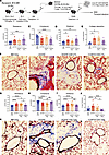 Figure 1
Day 28 CD8+ CAR19 T cells do not treat murine cGVHD/BOS, while CAR19 Tregs reduce disease. (A) B10.BR mice were conditioned with 120 mg/kg i.p. cyclophosphamide (day –3 and day –2) followed by 6.2 Gy total-body irradiation (day –1). On day 0, recipients received 107 T cell–depleted (TCD) bone marrow (BM) from hCD19tg F1 donors plus 71.5 × 10⁴ purified splenic T cells from C57BL/6 (B6)-Ly5 donors. Groups of cGVHD mice were infused with 0.5 × 10⁶ expanded CD8+ CAR19- or EGFR-control effectors on day 28, and on day 50 posttransplant mice were sacrificed for terminal analysis of cGVHD/BOS disease severity. (B–D) Pulmonary function test data from BM-only (n = 14), cGVHD (n = 9), EGFR CD8+ T cell–treated (n = 5), and CAR19 CD8+ T cell–treated (n = 11) mice including resistance (B), compliance (C), and elastance (D). (E and F) Quantification of collagen deposition (percentage area trichrome) from Masson’s trichrome–stained lung sections from CD8+ CAR19 T cell treatment studies (E) with representative images from BM-only (n = 5), cGVHD (n = 4), EGFR CD8+ T cell–treated (n = 4), and CAR19 CD8+ T cell–treated (n = 5) mice (F). (G–I) Pulmonary function test data from BM-only (n = 21), cGVHD (n = 17), EGFR CD4+ Treg–treated (n = 13), and CAR19 CD4+ Treg–treated (n = 17) mice including resistance (G), compliance (H), and elastance (I). (J and K) Quantification of collagen deposition (percentage area trichrome) from Masson’s trichrome–stained lung sections from CD4+ Treg treatment studies (J) with representative images from BM-only (n = 17), cGVHD (n = 11), EGFR CD4+ Treg–treated (n = 11), and CAR19 CD4+ Treg–treated (n = 14) mice (K).(F and K) Original magnification, ×200. (B–F) Data are pooled from 2 independent experiments. (G–K) Data are pooled from 4 independent experiments. Statistics shown are results of 1-way ANOVA with Tukey’s correction for multiple comparisons. *P < 0.05, **P < 0.01, ***P < 0.001, ****P < 0.0001.
Splenic CD8+ T cells were activated with anti-CD3/CD28 mAb–coated Dynabeads (15, 38) and transduced with hCAR19 or control (truncated EGFR) retrovirus. Transduced CD8+ T cells were uniformly enriched to greater than 90% EGFR purity. EGFR and CAR19 expression was confirmed on day 7 before administration (Supplemental Figure 2, A and B). Day 28 infusion of 0.5 × 10⁶ CAR19 or EGFR CD8+ T cells did not affect the >80% survival or weights that typically declined by ≤20% from pre–BM transplantation weights, although significant splenic lymphopenia was observed in comparison with cGVHD controls (Supplemental Figure 3, A–C). Day 28 CD8+ CAR19 T cells did not improve pulmonary function (Figure 1, B–D) despite significant peripheral B cell and GCB depletion in spleens and lungs (Supplemental Figure 3, D–F).
Spleen immunofluorescent staining on day 50, 22 days after day 28 infusion, showed that CD8+ CAR19 T cells decreased overall GC size, with more frequent, fragmented GCs lacking proper CD4+ T cell compartmentalization (Supplemental Figure 4, A–C). Splenocyte flow cytometry revealed no TFH, TFR, or TFR/TFH ratio changes (Supplemental Figure 3, G–I). Lung collagen deposition was not significantly reduced (Figure 1, E and F). Despite marked reduction in day 50 lung pathogenic IgG2c deposition (Supplemental Figure 5, A and B), improved clinical outcomes were not seen. Collectively, these data indicate that GCB depletion and reduced IgG2c deposition in the context of donor anti-host reactive allogeneic CD8+ CAR19 T cells are insufficient for cGVHD/BOS mitigation.
CAR19 CD4+ Tregs effectively treated murine cGVHD/BOS. Since CD8+ CAR19 T cells did not improve cGVHD/BOS, we explored CAR19 Tregs as an alternative therapy based on previous data demonstrating that CAR19 Tregs had a dual immune suppression and antigen-specific cytolytic capacity without detrimental immune responses inherent to CD8+ CAR19 T cells (15, 38). CAR19 Tregs were generated as described previously (15) using FoxP3-GFP reporter mice for flow sorting to 99% purity. Activated Tregs transduced with CAR19- or EGFR-only retrovirus were expanded with Dynabeads in IL-2, maintaining high FoxP3 purity (Supplemental Figure 2, C–E). The same suboptimal dose (0.5 × 10⁶) used for CD8+ CAR19 T cells was infused into day 28 cGVHD mice to intentionally minimize known beneficial effects with high-dose Tregs and facilitate detection of differences between groups (20, 21, 40).
Neither EGFR nor CAR19 Tregs adversely affected overall survival or weights (Supplemental Figure 6, A and B). In contrast to CD8+ EGFR or CAR19 T cells, CAR19 Tregs significantly improved pulmonary function across resistance, compliance, and elastance parameters (Figure 1, G–I) and significantly reduced lung collagen deposition in comparison with cGVHD controls and EGFR Treg–treated mice (Figure 1, J and K). CAR19 Treg–treated mice exhibited reduced pathology in the colon and liver, organs shown to be cGVHD target organs in this model (Supplemental Figure 7, A and B). However, lung histopathology scores were not reduced with either treatment. Possible explanations include the fact that histopathology scoring examines only a small fraction of lung tissue, whereas lung function measures the respiratory capacity of the lung as an entire organ, and lung injury repair, as assessed by histopathological scores, may require a longer time period than lung function to be readily detectable. These results demonstrate that CAR19 Tregs significantly alleviated murine cGVHD/BOS with multi-organ system disease.
CAR19 Treg–mediated treatment of cGVHD/BOS did not require GCB depletion. We sought to determine the mechanism(s) by which CAR19 Tregs improved cGVHD/BOS. Previously we reported that non-transplanted, day –1 cyclophosphamide-conditioned hCD19tgTg/0 mice experienced a profound and equivalent hCD19+ B cell depletion on day 5 after CAR19 Treg or CD8+ CAR19 T cell infusion; each had a potent effect on reducing B cell lymphoma (38). In an acute GVHD model, lethally irradiated hCD19tgTg/0 recipients of BALB/c (H-2d) donor BM with or without purified T cells with or without CD8+ EGFR or CAR19 T cells or EGFR Tregs died of GVHD with survival rates of 0%–20%, in contrast to 60%–100% survival in CAR19 Treg–treated recipients, most consistent with CAR19 Treg–mediated donor T cell alloresponse suppression (15). These data reflect CAR Treg cytolytic and suppressive properties, offering 2 possible mechanisms (B cell depletion and immunosuppression) for CAR19 Treg efficacy in cGVHD/BOS mice.
In cGVHD/BOS mice, CAR19 Treg infusion reduced TFH frequencies, increased TFR frequencies, and elevated TFR/TFH ratios in comparison with cGVHD controls (Figure 2, A–C). In contrast, EGFR Treg–treated cGVHD mice had TFH frequencies and TFR/TFH ratios similar to those of cGVHD controls. TFR frequencies were comparable to those of CAR19 Treg–treated mice, possibly reflecting Treg infusion. Although EGFR and CAR19 Treg spleen and lung frequencies and numbers were similar (Supplemental Figure 8, A–C), CAR19 Tregs increased TFRs by inhibiting cGVHD associated with reduced TFR frequencies or directly contributing GC TFRs (Figure 2D). These data are consistent with observations above that CAR19 Treg infusion augmented TFR/TFH ratios relative to EGFR Tregs (Figure 2C).
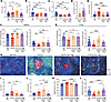 Figure 2
CAR19 Tregs inhibit GC reactions in murine cGVHD/BOS. (A–E) Day 50 spleens from cGVHD mice treated with EGFR or CAR19 Tregs were analyzed for frequency of TFHs (FoxP3–CXCR5+PD-1+BCL6+) of CD4+ (A), frequency of TFRs (FoxP3+CXCR5+PD-1+BCL6+) of CD4+ (B), TFH/TFR ratio (C), frequency of FoxP3(GFP)+ infused Tregs of TFRs (D), and frequency of GCBs of mouse CD19 positive (mCD19+) (E). (F and G) Frequency and counts of hCD19+ B cells of lymphocytes in day 50 spleen (F) and lung (G). (H) Representative images (original magnification, ×200) of spleen sections stained for GCs with peanut agglutinin (PNA; red), CD4 (green), and DAPI (blue). Scale bars: 200 μm. (I and J) Quantification of GC sizes (I) and frequencies per mm2 (J). (K and L) Day 50 spleens were analyzed for frequencies of mature B220–mCD19– plasma cells (PCs) of total CD138+BLIMP1+ PCs (K) or immature B220+mCD19+ PCs of total PCs (L). Data are pooled from 2–4 independent experiments. (A–C, E, and F) Day 50 spleens were analyzed from BM-only (n = 26), cGVHD (n = 25), EGFR CD4+ Treg–treated (n = 21), and CAR19 CD4+ Treg–treated (n = 23) mice. (D) Day 50 spleens were analyzed from EGFR CD4+ Treg–treated (n = 17) and CAR19 CD4+ Treg–treated (n = 25) mice. (G) Day 50 lungs were analyzed from BM-only (n = 17), cGVHD (n = 16), EGFR CD4+ Treg–treated (n = 14), and CAR19 CD4+ Treg–treated (n = 10) mice. (I and J) Day 50 spleens were analyzed from BM-only (n = 13), cGVHD (n = 8), EGFR CD4+ Treg–treated (n = 11), and CAR19 CD4+ Treg–treated (n = 10) mice. (K and L) Day 50 spleens were analyzed from BM-only (n = 10), cGVHD (n = 15), EGFR CD4+ Treg–treated (n = 13), and CAR19 CD4+ Treg–treated (n = 15) mice. Statistics shown are results of 1-way ANOVA with Tukey’s correction for multiple comparisons. *P < 0.05, **P < 0.01, ***P < 0.001, ****P < 0.0001.
Contrary to our expectation that CAR19 Tregs would more efficiently deplete GCBs compared with EGFR Tregs and be superior in treating cGVHD/BOS, CAR19 and EGFR Tregs similarly reduced GCB frequencies relative to cGVHD/BOS controls (Figure 2E). CAR19 and EGFR Tregs increased total splenic hCD19+ B cell frequencies and numbers to levels comparable to those in BM-only controls, consistent with Treg-mediated mitigation of cGVHD-induced B cell lymphopenia (Figure 2, F–H). However, CAR19 Tregs reduced the overall cGVHD-induced increase in GC size and frequency in comparison with cGVHD controls and EGFR Tregs (Figure 2, I and J).
Day 50 splenic plasma cell (PC) frequencies were analyzed as a possible source of high pathogenic antibody levels that might be differentially susceptible to CAR Treg–mediated suppression (Figure 2, K and L). CAR19 Tregs decreased mature PC frequencies and increased immature PC frequencies in comparison with cGVHD controls and EGFR Tregs, approaching BM-only control levels. Reduction of mature PCs was associated with significantly decreased lung pathogenic IgG2c deposition (Supplemental Figure 9, A and B).
Since we could not exclude rapid hCD19tgTg/0 B cell depletion by CAR19 Tregs that may have been missed on day 50, cGVHD/BOS recipients were studied on day 35, 7 days after EGFR or CAR19 Treg infusion, for evidence of hCD19tgTg/0 B cell depletion seen in cyclophosphamide-treated mice (38). Unexpectedly, no measurable B cell depletion was observed with CAR or EGFR Tregs in spleen or lungs (data not shown). Specifically, there were no frequency or count differences between CAR19 and EGFR Tregs in these tissues early post-infusion (data not shown). Together, these findings suggest that CAR19 Tregs suppress pathological GC reactions without depleting B cells, resulting in reduced cGVHD/BOS, enhanced B cell recovery, and prevention of secondary lymphoid organ involution typically seen in untreated cGVHD recipients.
CAR19 Tregs maintained cytolytic capacity in vitro and in cyclophosphamide-treated syngeneic recipients. To better understand why CAR19 Tregs did not deplete B cells in cGVHD/BOS, we reevaluated CAR19 Treg cytolytic potential against B cells. We cocultured EGFR or CAR19 Tregs and CD8+ T cells for 5–48 hours with hCD19tgTg/0 B cells (Figure 3, A–E, and Supplemental Figures 10–12). CAR19 Tregs cocultured with and activated by hCD19tgTg/0 B cells exhibited higher baseline CD107a and perforin expression compared with EGFR Tregs, and upregulated CD107a, FasL, perforin, granzyme A (but not granzyme B), TNF-α, and IFN-γ upon CAR stimulation, albeit at lower levels than CD8+ CAR19 T cells. These results confirm that CAR19 Tregs possess an antigen-specific cytolytic capacity with a distinct effector profile.
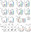 Figure 3
CAR19 Tregs maintain cytolytic capacity in vitro and in cyclophosphamide-treated syngeneic recipients. (A–E) Frequency and MFI of cytolytic markers after EGFR CD4+ Treg (n = 3), CAR19 CD4+ Treg (n = 3), EGFR CD8+ cytotoxic T lymphocyte (CTL) (n = 3), and CAR19 CD8+ CTL (n = 3) coculture with freshly isolated hCD19+ B cells at 1:5 effector/B cell ratio for 5–48 hours: (A) CD107a (5 hours), (B) FasL (48 hours), (C) perforin (24 hours), (D) granzyme A (48 hours), (E) TNF-α (24 hours). (F) Forty-eight-hour in vitro flow killing assay of freshly isolated hCD19tgTg/0 or wild-type (WT) B cells cocultured with EGFR CD4+ Tregs (n = 4), CAR19 CD4+ Tregs (n = 4), EGFR CD8+ CTLs (n = 4), and CAR19 CD8+ CTLs (n = 4) at a ratio of 5 effectors to 1 target with addition of 5 μg/mL αIgM and 100 ng/mL BAFF. (G and H) cGVHD/BOS mice were set up as previously described and infused with 3.0 × 10⁶ effectors on day 28 (Figure 1A). Syngeneic hCD19tgTg/0 mice were treated with 300 mg/kg cyclophosphamide the day before infusion of 3.0 × 10⁶ effectors. Spleen and lung tissues were harvested 5 days after infusion for flow analysis. Representative flow cytometry plots of hCD19+ B cells of lymphocytes from spleen tissues (G) with bar graph summary of spleen (left) and lung (right) tissues (H) from in vivo killing experiment in control (no treatment) (n = 3), CAR19 CD8+ CTL–treated (n = 3), EGFR CD4+ Treg–treated (n = 3), and CAR19 CD4+ Treg–treated (n = 3) mice. Data are representative of 2–3 independent experiments. Statistics shown are results of 1-way ANOVA with Tukey’s correction for multiple comparisons. *P < 0.05, **P < 0.01, ***P < 0.001, ****P < 0.0001.
To quantify CAR19 Treg–mediated B cell cytolysis, we performed a flow cytometry–based killing assay by coculturing naive hCD19+ or wild-type B cells with EGFR or CAR19 Tregs at a 5:1 ratio of Tregs to B cells for 48 hours. Controls were CD8+ EGFR or CAR19 CD8+ T cells. B cells were activated with anti-IgM and B cell–activating factor (BAFF). CAR19 Tregs and CD8+ CAR19 T cells demonstrated robust killing of hCD19+ B cells, with an average of 1.3% and 0.85% live B cells remaining, respectively, compared with 39.1% in control cultures without effectors (Figure 3F). CAR19 Treg killing was hCD19 antigen specific with no cytolysis observed against wild-type B cells; EGFR Tregs showed no cytolysis under any condition.
To assess in vivo cytolytic capacity, hCD19tgTg/0 recipients were pretreated with cyclophosphamide, infused with 3 × 10⁶ syngeneic CAR19 Tregs or CD8+ CAR19 T cells (38), and set up in parallel with the cGVHD/BOS model. This ensured that day 28 cGVHD CAR19 Treg or CD8+ CAR19 T cell infusion coincided with day 0 infusion of the same cell aliquot given to non-transplanted, cyclophosphamide-treated mice (38). By day 5, splenic hCD19+ B cell frequencies decreased from 16.0% in non-infused mice to 0.20% and 2.93% in cyclophosphamide-treated mice receiving CD8+ CAR19 T cells and CAR19 Tregs, respectively, with similar lung B cell aplasia (Figure 3, G and H). In contrast, in cGVHD/BOS mice, only CD8+ CAR19 T cells reduced splenic hCD19+ B cell frequencies (from 24.3% to 7.2%), whereas CAR19 Tregs had no effect. These results demonstrate that the same CAR19 Treg aliquot retained B cell elimination capacity in vitro and in cyclophosphamide-treated, non–BM transplantation recipients but not in cGVHD/BOS mice.
CAR19 Treg cytolysis was uninhibited by cGVHD serum and required cell-cell contact. Next, in order to determine whether GCBs from cGVHD/BOS were resistant to CAR19 Treg cytolysis, or whether soluble factors in the in vivo cGVHD environment might account for the lack of profound CAR19 Treg–mediated B cell killing, a 48-hour flow cytometry–based killing assay was used. B cells isolated from day 28 cGVHD/BOS mice were cocultured at a 5:1 Treg/B cell ratio, with or without serum from day 28 BM-only or cGVHD /BOS mice (1:10 dilution). Adding cGVHD serum significantly increased baseline survival of ex vivo cGVHD B cells compared with no serum or BM-only serum (Figure 4A). CAR19 Tregs still mediated significant killing of ex vivo cGVHD B cells, with an average of 3.6% live B cells remaining compared with 13.0% in control cultures without effectors (Figure 4B). EGFR Tregs showed no cytolytic activity. Importantly, CAR19 Tregs maintained robust cytolysis in the presence of BM-only and cGVHD serum (Figure 4, C and D), with no significant difference between serum groups (Figure 4E).
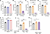 Figure 4
CAR19 Treg cytolytic activity is uninhibited by cGVHD/BOS serum and requires cell-cell contact. (A–G) All in vitro killing assays were cultured for 48 hours with ex vivo day 28 cGVHD/BOS B cells at an effector/target ratio of 5:1. (A) Frequency of live cGVHD B cells in target-only cultures of the killing assay following 1:10 addition of BM-only or cGVHD serum. (B–D) In vitro flow killing assay of cGVHD B cells by CAR19 versus EGFR Tregs with no serum (B) or 1:10 serum addition from BM-only mice (C) or cGVHD/BOS mice (D). (E) Frequency of CAR19 Treg killing of cGVHD B cells from B–D. (F and G) In vitro flow killing assay in a Transwell plate with freshly isolated hCD19tgTg/0 B cells and addition of 5 μg/mL αIgM and 100 ng/mL BAFF. hCD19+ B cells were added to lower wells. Effectors were added to the lower wells (F) or upper inserts (G). Data are representative of 3 independent experiments. (A–F) Groups were analyzed from B cell–only (n = 4), EGFR CD4+ Treg (n = 4), and CAR19 CD4+ Treg (n = 4) cocultures. (G) Groups were analyzed from B cell–only (n = 4), EGFR CD4+ Treg (n = 3), and CAR19 CD4+ Treg (n = 3) cocultures. Statistics shown are results of 1-way ANOVA with Tukey’s correction for multiple comparisons. *P < 0.05, **P < 0.01, ***P < 0.001, ****P < 0.0001.
We investigated whether in vitro CAR19 Treg cytolysis required direct cell-cell contact. Using Transwell plates, hCD19tgTg/0 naive B cells were plated in the lower chambers with effectors in the same chamber or separated by a membrane at 5:1 effector/B cell ratios. Robust cytolysis was observed only when CAR19 Tregs or CD8+ CAR19 T cells were in direct contact with B cells; no killing occurred when separated, and B cell survival increased (Figure 4, F and G), indicating that CAR19 Treg cytolysis is contact dependent. Together, these findings indicate that CAR19 Tregs are fully cytolytic via contact-dependent mechanisms, and neither cGVHD B cell resistance nor soluble factors in cGVHD/BOS serum inhibited CAR19 Treg cytolysis ex vivo.
CAR19 Tregs directly interacted with B cells and inhibited B cell activation in cGVHD/BOS. To better understand the mechanism(s) by which CAR19 Tregs treat cGVHD/BOS, we used spatial proteomics to deconvolute cell-cell interactions in cGVHD/BOS (Supplemental Figure 13A). On day 33, 5 days after Treg infusion into cGVHD/BOS mice, spatial proteomics identified distinct Treg localization patterns between mice given EGFR and CAR19 Tregs. Compared with mice given EGFR Tregs, spleens from mice given CAR19 Tregs demonstrated a higher infused Treg proportion surrounding B cell follicles, based on individual cell type deconvolution and annotation (Supplemental Figure 13, B and C) and spatial domain analysis (Figure 5A). B cell subtyping within B cell zones revealed an approximately 1.5-fold decrease in the proportion of CD40+ (activated) B cells in CAR19 compared with EGFR Treg–treated mice, with a reciprocal approximately 1.5-fold increase in naive (non-activated) B cells (Figure 5B). Of CD40+ B cells, the mean intensity of CD40 expression was about 25% greater in EGFR Treg mice compared with CAR19 Treg mice (Figure 5C).
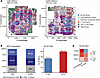 Figure 5
Day 28 CAR19 Tregs predominantly localize to follicular T cell–rich areas instead of B cell–rich areas in cGVHD/BOS spleens. (A) Annotated spatial domains detected using local indicators of spatial association (LISA) functions for each cell at multiple radii followed by manual annotation based on cell type distribution patterns. (B) Proportions of B cells versus activated B cells in B cell follicle spatial domain. Left: Frequencies of total cells in domains. Right: Direct comparison of activated B cell frequency. BCZ, B cell zone. (C) Heatmap of B cell–associated marker mean intensity differences between CAR19 and EGFR Treg–treated mice. Color maps to fold change between conditions. Increased fold change (red) was seen for most markers in EGFR samples. The left column is fold change difference by marker in activated B cells, while the right column is for B cells. Data analysis was performed on a single slide for each condition.
Visualization of direct CAR19 Treg and B cell colocalization was not observed, possibly reflecting earlier or transient effects. Therefore, we addressed larger spatial interactivity patterns by evaluating splenic B cell follicle and Treg-enriched spatial zone boundary regions (Figure 6A), serving as a proxy for interaction opportunities between these cell types. Compared with EGFR Treg mice, CAR19 Treg mice had approximately 2-fold as many boundaries with an approximately 1.5-fold greater shared interaction length between B cell follicles and Treg-enriched zones (Figure 6B). These spatial proteomics results demonstrate that CAR19 Treg localization is distinct from EGFR Tregs, and that the splenic architecture in CAR19 Treg–treated mice would provide more opportunities for CD19+ B cell interaction at the B cell follicle boundary zones. These interactions may drive Treg-mediated suppression of B cell activation.
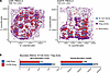 Figure 6
CAR19 Treg–enriched zones show greater interaction boundaries with follicular B cell zones than EGFR Treg controls. (A) Secondary annotation of B cell zone and Treg zone spatial domains from Figure 5 with inclusion of detected boundaries of interaction between them. (B) Quantifications of B cell and Treg zone boundaries of interaction. Left: Number of individual boundaries detected. Right: Total combined boundary length in pixels. Data analysis was performed on a single slide for each condition.
CAR19 Tregs exhibited a more robust suppressive profile compared with EGFR Tregs. With perceived CAR19 Treg–B cell interaction increases, we investigated whether such interactions may render CAR19 Tregs more suppressive after CD19 engagement. We cocultured CAR19 or EGFR Tregs with hCD19tgTg/0 naive B cells at a 1:5 Treg/B cell ratio for 5–48 hours (Figure 7, A–D, and Supplemental Figure 14). Notably, CAR19 Tregs exhibited higher baseline PD-1 and TIGIT expression compared with EGFR Tregs. Upon CAR stimulation by hCD19+ B cells, CAR19 Tregs showed increased frequencies and mean fluorescence intensities (MFIs) for molecules associated with suppression, including IL-10, PD-1, and TIGIT, and elevated CTLA-4 MFI relative to EGFR Tregs.
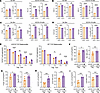 Figure 7
CAR19 Tregs have greater suppressive capacity compared with EGFR Tregs in vitro and in vivo during murine cGVHD/BOS. (A–D) Frequency and MFI of suppressive markers expressed by EGFR CD4+ Tregs (n = 3) or CAR19 CD4+ Tregs (n = 3) after 48 hours of no stimulation or coculture with hCD19tgTg/0 B cells: (A) IL-10, (B) PD-1, (C) Tigit, (D) CTLA-4. (E) In vitro suppression of CD4+ T cell proliferation by EGFR Tregs (n = 4) or CAR19 Tregs (n = 4) cocultured with hCD19+ TCD splenocytes (left) or wild-type TCD splenocytes (right). (F–I) Frequency and MFI of infused FoxP3(GFP)+ Tregs from day 50 spleens of cGVHD mice treated with EGFR Tregs (n = 4) or CAR19 Tregs (n = 6) expressing PD-1 (F), Tigit (G), CD127 (H), and LAG3 (I). Data are representative of 3 independent experiments. Statistics shown are results of 1-way ANOVA with Tukey’s correction (A–D and F–I) or 2-way ANOVA with Šidák’s correction for multiple comparisons (E). *P < 0.05, **P < 0.01, ***P < 0.001, ****P < 0.0001.
In vitro suppression assays were performed to compare CAR19 and EGFR Treg potency. Treg groups were cocultured at varying ratios with congenic conventional T cells (Tcons) labeled with CellTrace CFSE proliferation dye. TCD splenocytes from hCD19tgTg/0 or wild-type B6 mice were added at 1:1 with Tcons. CAR19 Tregs demonstrated superior suppression of T cell proliferation compared with EGFR Tregs, with more consistent and greater suppression when cultured with hCD19+ TCD splenocytes (Figure 7E).
Flow cytometry analysis of day 50 cGVHD/BOS spleens revealed that day 28 CAR19 Treg infusion exhibited a more suppressive and memory-like phenotype compared with EGFR Tregs, indicated by increased frequencies and MFIs of TIGIT, PD-1, LAG3, CD127, and KLRG1 (Figure 7, F–I, and Supplemental Figure 15).
Collectively, these findings highlight that CAR19 Tregs are more suppressive than EGFR Tregs in vitro and in vivo, and that their suppressive phenotypes can be effectively mediated through CAR signaling.


