
Oncology
Anti-Notch plus endocrine therapy in ER+ breast cancer inhibits cancer stem cells by increasing DAXX
Abstract
Resistance to endocrine therapy (ET) in ER+ breast cancer is mediated by Notch signaling, but the clinical application of anti-Notch therapy has been limited by the lack of predictive biomarkers. To identify Notch-regulated biomarkers, we conducted a pre-surgical window study evaluating ET combined with the γ-secretase inhibitor (GSI) MK-0752. RNA expressions in tumors were measured using an Affymetrix array and by real-time PCR. ET plus GSI showed more genes were decreased than ET alone. Specifically, DAXX, NOXA, and LFNG RNAs were increased, while fifteen additional transcripts were decreased. Mechanistically, GSI reduced Notch1 occupancy at CSL-binding elements within HES1, HEY2, HEYL, CCND1, MKI67, and DAXX genes, and inhibited cancer stem cells (CSCs) by 90% to 100%. This anti-CSC effect required DAXX, while GSI treatment or Notch1/4 knockdown increased DAXX expression, suggesting transcriptional repression by Notch. Using mouse tumor xenograft studies, ET plus MK-0752 resulted in complete regression of MCF-7 tumors, with DAXX-high tumors showing greater treatment sensitivity. Clinically, high DAXX expression was associated with improved recurrence-free and overall survival. This study found that anti-Notch plus ET in ER+ breast cancer inhibits cancer stem cells by increasing DAXX, a promising predictive biomarker. These findings support clinical evaluation of therapies that increase DAXX expression.
Authors
Kathy S. Albain, Debra Wyatt, Andrei Zlobin, Susan G. Hilsenbeck, Cheryl M. Czerlanis, Daniel S. Peiffer, Kyle R. Convington, Constantine Godellas, Shelly S. Lo, Patricia A. Robinson, Kathy Czaplicki, Barbara Busby, Davide Bova, Ping Tang, Patrick J. Stiff, Suzanne A.W. Fuqua, Lucio Miele, Clodia Osipo
Abstract
BACKGROUND. Hepatocellular carcinoma (HCC) exhibits molecular heterogeneity that challenges histopathologic classification and biomarker discovery. We assessed whether spatially resolved N-glycan imaging with machine learning could classify tumor regions and infer glutamine synthetase (GS) status. METHODS. In this retrospective study, MALDI mass spectrometry imaging of N-glycans was performed on formalin-fixed, paraffin-embedded sections from two independent cohorts (discovery, n = 88; validation, n = 60) with pathologist annotation. An XGBoost classifier was trained on 90 discriminative N-glycan features using patient-grouped cross-validation. Performance was assessed by AUC for pixel- and biopsy-level discrimination of tumor from adjacent non-tumor tissue, and for GS status classification. RESULTS. Pixel-level AUCs were 0.95 (cross-validation) and 0.89 (external validation); biopsy-level AUCs were 1.0 and 0.97, correctly identifying 97% of tumor-containing biopsies. Probability maps recapitulated pathologist-defined boundaries; UMAP embeddings captured inter- and intratumoral heterogeneity. Discriminative species (m/z 2393.846, 1905.634, 1743.579, 1809.639) reflected complex, fucosylated, branched remodeling. N-glycans bearing six GlcNAc residues were enriched in GS+ (n = 45) versus GS− (n = 17) tumors (P = 0.001) and discriminated GS status (AUC = 0.75), consistent with GLUL and MGAT5 upregulation in TCGA-LIHC. CONCLUSION. MALDI N-glycan imaging with machine learning enables spatially resolved, objective classification of HCC and links glycan phenotypes to tumor-associated metabolic programs. TRIAL REGISTRATION. Not applicable; retrospective analysis of archival, de-identified tissue. FUNDING. NIH/NCI R01CA285370, 1R01CA289381, R33CA267226, R01CA282022, R21CA263464, R21CA286287, R01CA253460, S10OD030212, R01CA251155, R01CA250227, U01CA271887, P50CA295495, P30CA138313, P20GM130457, P30DK123704, P30DK120531,R24DK139775; NIH/NIA R01AG078702; Smart State Endowment, State of South Carolina; LeDucq Foundation.
Authors
Muhammed F. Bayram, Jade K. Macdonald, Andrew DelaCourt, Peggi M. Angel, Richard R. Drake, Aatur Singhi, David Geller, Satdarshan P. Monga, Amit Singal, Anand Mehta
Abstract
BACKGROUND. This prospective, single-arm, multicenter phase 1/2 trial evaluated vorinostat added to standard graft-versus-host disease (GVHD) prophylaxis in pediatric, adolescent, and young adult (AYA) patients undergoing allogeneic hematopoietic cell transplantation (HCT) from HLA-matched related, HLA-matched unrelated, and haploidentical donors. METHODS. Patients aged 3–39 years received twice-daily vorinostat with tacrolimus/methotrexate after HLA-matched HCT from day −10 to +30, or with post-transplant cyclophosphamide/tacrolimus/mycophenolate mofetil after haploidentical HCT from day +5 to +30. The primary endpoint was cumulative incidence of grade II–IV acute GVHD by day +100. All outcomes were based on intention-to-treat analysis. RESULTS. Forty-three patients were enrolled; median age was 19 years, with 74% receiving HLA-matched and 26% haploidentical HCT. The recommended phase 2 dose was 60 mg/m² twice daily. No dose-limiting toxicities, unexpected vorinostat-related serious adverse events, or primary graft failures occurred. Median neutrophil and platelet recovery occurred at 14 and 18 days, respectively. Day +100 grade II–IV and III–IV acute GVHD were 14% (95% CI, 5.6, 26) and 4.7% (95% CI, 0.83, 14), respectively. One-year overall survival was 88.4% (95% CI, 79.3, 98.5), relapse 14% (95% CI: 5.6, 26), nonrelapse mortality 4.7% (95% CI: 0.8, 14), and GVHD-free/relapse-free survival 55.8% (95% CI: 42.8, 72.8). Correlative studies demonstrated on-target HDAC activity, with increased histone acetylation and lower proinflammatory cytokines. CONCLUSION. These findings support randomized evaluation of vorinostat-based GVHD prophylaxis in pediatric and AYA HCT. TRIAL REGISTRY. ClinicalTrials.gov, NCT03842696.
Authors
Nicolas Parnell, Xiao Cao, Jan H. Beumer, Thomas M. Braun, Yilei Cui, Gary J. Fisher, Guoqing Hou, Julianne Holleran, Tracey Churay, Michelle Rozwadowski, Luna Heider, Mark T. Vander Lugt, Carrie L. Kitko, April L. Rahrig, Ed Peres, Kirsten M. Williams, Julie-An Talano, Vanessa A. Fabrizio, Ghada Abusin, Gregory A. Yanik, John Magenau, Mary Riwes, Marcus J. Geer, Sarah Anand, Monalisa Ghosh, Attaphol Pawarode, Kristen Votruba, Pavan Reddy, Sung Won Choi
Abstract
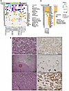
Metastases in renal cell carcinoma (RCC) typically arise from large primary tumors. However, a subset of patients with small renal masses (SRMs; ≤4 cm) can develop metastatic disease. Identifying these tumors is clinically important, as many SRMs are managed with active surveillance, and their study may provide insight into the early acquisition of metastatic competence. It remains unclear whether these tumors acquire distinct metastatic programs or instead show premature activation of the same aggressive programs typically associated with larger tumors. Here, we performed integrated morphological and molecular profiling of a multiinstitutional cohort of metastatic SRMs, including whole-exome sequencing and RNA-Seq, using nonmetastatic primary tumors as controls. Among metastatic, non–clear cell SRMs, we identified NF2-altered tumors, ELOC-mutated RCC, and an mTOR-driven eosinophilic vacuolated tumor. Metastatic clear cell SRMs were enriched by multi-hit aggressive genotypes, including recurrent losses of chromosomes 8p, 9, and 14q, as well as co-occurring driver alterations (≥2 events), including BAP1 and mTOR pathway genes. Transcriptomic analyses revealed enrichment of the non-negative matrix factorization 3 (NMF3) subtype from the IMmotion151 trial-based taxonomy, along with metabolic rewiring and reduced cytotoxic immune effector function. Collectively, these findings identify molecular programs associated with metastatic competence in SRMs, highlight the importance of genomic studies of equivocal non-clear cell SRMs, and provide a biological framework for risk stratification in patients often considered for active surveillance.
Authors
Payal Kapur, Daria Beshnova, Hua Zhong, Ruby Sharma, Pooja Ghatalia, Angela Yoo, Daniel D. Le, Ratna Mukhopadhyay, Alana Christie, Jeffrey Miyata, Shuanzeng Wei, Rana R. McKay, Dinesh Rakheja, Satwik Rajaram, Robert G. Uzzo, A. Ari Hakimi, Zora Modrusan, James Brugarolas
Abstract
Despite advances in treatment approaches for lung cancer, the morbidity and survival of lung cancer patients with malignant pleural effusions (MPE) remain poor. This is in part due to gaps in understanding the role of immune cells in the pleural fluid microenvironment. We performed single cell analysis with flow cytometry validation of CD45+ cells in eight malignant and five benign pleural fluid (BPE) specimens to identify changes in the transcriptomic landscape of immune cells across disease states. We found upregulation of pro-inflammatory signaling pathways, including interferon and TNF signaling, in T cells, B cells, and macrophages in benign compared to malignant pleural effusions. Pro-inflammatory HLA-DR+ macrophages were associated with good survival outcomes while pro-tumorigenic HLA-DR- macrophages with upregulation of angiogenesis, TGFβ, and fibronectin signaling were associated with poor survival outcomes in patients with MPE. We also validated these findings with macrophage cell surface expression markers using flow cytometry in 14 MPE and 7 BPE specimens. Finally, we performed multiplex cytokine analysis which showed enrichment of the type 3 inflammatory cytokine, IL17A, in MPE as a putative mechanism for macrophage reprogramming. These data provide a rich resource for interrogating the immune cell types and states present across the spectrum of pleural disease. They offer not only prognostic value for patient outcomes at the time of pleural fluid collection, but also insights into novel immunotherapy targets.
Authors
Aaditya Khatri, Huimin Wang, Zhicheng Ji, Prekshaben Patel, Smita K. Nair, Javid P. Mohammed, Beth H. Shaz, Andrew B. Nixon, Scott M. Palmer, Kamran Mahmood
Endothelial cell cycle inhibition enables blood vessel maturation to normalize the tumor vasculature
Abstract
Dysfunctional tumor vessels promote disease progression, whereas improved function enhances therapeutic delivery. However, current approaches to normalize tumor vasculature have limited efficacy. In vascular malformations, vessels are similarly dysfunctional, with endothelial cell (EC) hyperproliferation impairing arterial-venous specification. These defects are corrected with palbociclib, a cyclin-dependent kinase 4/6 inhibitor (CDK4/6i) that has beneficial effects on tumor and immune cells, but the effects on tumor vasculature are not well characterized. In our studies, murine mammary tumor ECs (TECs) exhibited disrupted cell cycle and specification, and CDK4/6i promoted TEC cycle control, enabling improved tumor vascular function. To investigate transcriptomic changes, we performed single-cell RNA sequencing (scRNAseq) of treated and untreated tumors, and healthy tissues. CDK4/6i-mediated TEC cycle arrest promoted arterial-venous specification, cellular junctions, and pericyte association, and suppressed glycolytic and immunosuppressive gene expression. These effects were associated with increased vessel perfusion, decreased tumor hypoxia, and a more favorable immune landscape with immunotherapy. In scRNAseq datasets from patients treated long-term with CDK4/6i, TECs exhibited similar transcriptomic changes associated with arterial-venous specification, pericyte recruitment, and immune signaling. Thus, in contrast to current strategies, CDK4/6i-mediated vascular changes may be maintained with continued treatment, highlighting the relevance of modulating TEC cycle to improve vessel maturation/function.
Authors
Shelby R. Cain, Gael Genet, Nafiisha Genet, Jordon W. Aragon, Madeline G. Jackson, Victoria M. Milosek, Mark R. Schwartz, Umadevi Paila, Aleksandra Cwiek, Zaneta Markowska, Nicholas W. Chavkin, Richard J. Price, Andrew C. Dudley, Karen K. Hirschi
Abstract
Wnt signaling drives tumorigenesis in multiple cancers, in part through complex interactions with other oncogenic pathways including the MAPK cascade. In Wnt-addicted cancers, pharmacologic and genetic inhibition of Wnt signaling activates multiple receptor tyrosine kinases (RTKs), increases ERK phosphorylation and induces MAPK target gene expression, but the specific RTKs responsible for this MAPK hyperactivation are not known. Here we performed phosphotyrosine-targeted mass spectrometry, which revealed robust phosphorylation of EPHA2 and EGFR upon Wnt inhibition. Unexpectedly, we find that in xenografts, EPHA2 suppresses EGFR and ERK activation. Most notably, the increased ERK phosphorylation observed in EPHA2 KO tumors is transcriptionally inert, as there is no concomitant increase in MAPK target gene expression until concomitant Wnt inhibition. This suggests a Wnt-activated transcriptional repressor such as GATA3 that gates MAPK signaling in Wnt-high cancers. While Wnt-high KRAS-mutant cancers are resistant to erlotinib alone, adding Wnt inhibitor mitigates this resistance. Additionally, loss of EPHA2 enhances their sensitivity to both erlotinib and Wnt inhibitors. These studies therefore identify therapeutic vulnerabilities in Wnt-high tumors, even within traditionally EGFR inhibitor-resistant, RAS-mutant contexts.
Authors
Shawn R. Wadia, Changyuan Hu, Siddhi Patnaik, Shreya Sridharan, Roger J. Daly, David M. Virshup, Babita Madan
Abstract
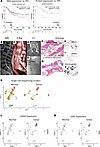
Estrogen can promote aggressive tumor phenotypes in estrogen receptor–positive (ER+) breast cancer; however, ER– cell lines are not widely considered estrogen responsive. Noncanonical estrogen-stimulated pathways such as the membrane-bound G protein–coupled estrogen receptor (GPR30) can mediate migratory and proliferative phenotypes in breast cancer and are postulated to promote resistance to aromatase therapies. Moreover, dysregulation of UDP-glucose 6-dehydrogenase (UGDH), a ubiquitously expressed enzyme critical to the metabolism of UDP-glucuronic acid into extracellular matrix precursors and hormone regulation, is associated with tumorigenesis. Here, we illustrated the impact of estrogen stimulation on tumor phenotypes in ER+ and ER– cell models in vitro and in vivo. We then demonstrated UGDH’s association with metastatic breast cancer via single-cell sequencing of patient specimens. Genetic knockdown of UGDH blunted estrogen-stimulated tumor phenotypes in vitro, ex vivo, and in vivo using both ER+ and ER– breast cancer lines. Finally, we demonstrated that UGDH knockdown blunted noncanonical estrogen stimulation through GPR30. Ultimately, our study validated prior studies demonstrating estrogen-responsive malignant phenotypes in ER– breast cancer and demonstrated that estrogen-stimulated breast cancer progression can be mediated through noncanonical pathways (e.g., UGDH/GPR30), regardless of ER status.
Authors
Meghan J. Price, Annee D. Nguyen, Corinne H. Strawser, Trupti Trivedi, Cesar C.D. Baeta, Catherine Lavau, Jovita K. Byemerwa, Debarati Mukherjee, Suzanne E. Wardell, Sandeep Artham, Vardhman Kumar, Shyni Varghese, C. Rory. Goodwin

Abstract
BACKGROUND. Loss of the Y chromosome (LOY) is a frequent event in male tumors and has been linked to cancer progression. However, the degree of mosaic LOY (mLOY) within normal tissues from men with or without cancer remains uncharacterized. METHODS. Here we used a FISH-based assay targeting X- and Y-chromosome centromeres to perform a pan-organ analysis of mLOY in 1,000 male tissue samples from 405 individuals representing 11 organs. Automated image processing generated a quantitative FISH-based mLOY score (YchrFISH) that we validated against a transcriptomic surrogate of Y-chromosome dosage from RNA-seq data. RESULTS. mLOY burden varied by tumor type, with highest degree in colorectal carcinoma. Across tissue groups, YchrFISH scores declined progressively from normal tissues of cancer-free men to histologically normal tissues adjacent to cancer and carcinoma (P < 0.0001). Paired analyses confirmed consistently greater mLOY in malignant compared with tumor-adjacent histologically normal tissue in different organs. Spatially resolved RNA-seq maps of bladders removed for cancer demonstrated a transcriptional gradient of Y-chromosome loss from normal urothelium through intraepithelial neoplasia to invasive carcinoma. CONCLUSION. mLOY gradients exist across histologically normal and malignant tissues, consistent with the concept of field cancerization. Our findings support epithelial mLOY as a biomarker of early malignant transformation and, to our knowledge, a previously unrecognized hallmark of male oncogenesis. FUNDING. NIH grants R35CA294022, P01CA163227, and P50CA97186 (the Pacific Northwest Prostate Cancer SPORE) and the Institute for Prostate Cancer Research.
Authors
Arkadiusz Gertych, Huihui Ye, Xingyu Chen, Eric Vail, V. Krishnan Ramanujan, Lauren Brady, Lawrence D. True, Peter S. Nelson, Peter R. Carroll, Dan Theodorescu
Abstract
Menin is a scaffolding protein that interacts with context-specific partners to regulate gene expression. In MLL-rearranged leukemias, Menin:MLL interactions drive leukemogenesis and Menin inhibitors have been FDA approved for these cancers. We previously reported that Menin promotes oncogenic phenotypes in Ewing sarcoma (EwS). Here, we sought to define EwS-specific functions of Menin and determine if Menin inhibitors could be therapeutically leveraged for these tumors. Genetic knockout of Menin had no impact on EwS cell proliferation in vitro, but metastatic potential of Menin-depleted cells in vivo was impaired. Transcriptional profiling of Menin knockout cells in vitro showed reproducible downregulation of MYC signature genes and upregulation of developmental programs. Conversely, transcriptional rewiring of developmental genes and restoration of MYC target gene expression were evident in tumors that arose from Menin knockout cells. Exposing EwS cells to the Menin inhibitor VTP50469 (revumenib) inhibited expression of MYC targets and co-immunoprecipitation studies detected Menin:MYC interactions that were partially disrupted by the drug. Metastatic colonization of disseminated EwS cells in vivo was significantly inhibited in mice fed VTP50469 chow. Together these findings implicate Menin as a mediator of EwS metastasis and suggest that Menin inhibitors warrant investigation as novel therapeutics for patients with high-risk disease.
Authors
Katherine A. Braun, Nicolas M. Garcia, Mohamed A. Ahmed, Darleen S. Tu, Stephanie I. Walter, Emma D. Wrenn, Megan E. B. Dean, Neerja Katiyar, Elizabeth R. Lawlor
No posts were found with this tag.


