
Neuroscience

Abstract
In multiple sclerosis (MS) lesions, CD8 T cells outnumber CD4 T cells, suggesting that they contribute to MS pathology. However, little is known about the role of CD8 T cells in MS, partly due to the prevalent use of experimental autoimmune encephalomyelitis (EAE) models mediated by CD4 T cells, which have limited involvement of CD8 T cells. Importantly, MS and EAE differ in both their distribution of CNS lesions and neurologic deficits, indicating differences in CNS inflammation. MS lesions are more commonly found in the brain, whereas EAE lesions are more frequent in the spinal cord. Additionally, neurologic deficits in MS rarely parallel the ascending paralysis typical for CD4 T cell–mediated EAE (CD4-EAE). In contrast, CD8-EAE models suggest that CD8 T cells preferentially cause brain inflammation; however, little is known about how brain and spinal cord inflammation may differ, or how CD8 T cells contribute to these differences. We have established an adoptive CD8-EAE mouse model characterized by brain-centered inflammation, ataxia, and weight loss. CNS inflammation in the brain and spinal cord differed in immune cell numbers, cellular composition, and inflammatory signatures. CD8-EAE could be suppressed by blocking IFN-γ, and exacerbated by blocking PD-1, with concomitant changes in the numbers of CNS-infiltrating monocytes. Most CD8 T cells in the CNS were CD11c+, suggesting that they are the pathogenic subset. We describe a robust CD8-EAE model, identify differences between brain and spinal cord inflammation, and characterize mechanisms that control CD8 T cell–mediated neuroinflammation, thereby furthering understanding of EAE and MS.
Authors
Daniel Hwang, Gholamreza Azizi, Larissa Lumi Watanabe Ishikawa, Maryam Seyedsadr, Arin Cox, Soohwa Jang, Ezgi Kasimoglu, Abdolmohamad Rostami, Guang-Xian Zhang, Bogoljub Ciric
Abstract
Biallelic loss-of-function variants in the adaptor protein complex 4 (AP-4) disrupt trafficking of transmembrane proteins at the trans-Golgi network, including the autophagy-related protein 9A (ATG9A), leading to childhood-onset hereditary spastic paraplegia (AP-4-HSP). AP-4-HSP is characterized by features of both a neurodevelopmental and degenerative neurological disease. To investigate the molecular mechanisms underlying AP-4-HSP and identify potential therapeutic targets, we conducted an arrayed CRISPR/Cas9 loss-of-function screen of 8,478 genes, targeting the ‘druggable genome’, in a human neuronal model of AP-4 deficiency. Through this phenotypic screen and subsequent experiments, key modulators of ATG9A trafficking were identified, and complementary pathway analyses provided insights into the regulatory landscape of ATG9A transport. Knockdown of ANPEP and NPM1 enhanced ATG9A availability outside the trans-Golgi network, suggesting they regulate ATG9A localization. These findings deepen our understanding of ATG9A trafficking in the context of AP-4 deficiency and offer a framework for the development of targeted interventions for AP-4-HSP.
Authors
Marvin Ziegler, Cedric Günter, Julian E. Alecu, Xutong Xue, Hyo-Min Kim, Afshin Saffari, Alexandra K. Davies, Mustafa Sahin, Darius Ebrahimi-Fakhari
Abstract
Skeletal muscle pathology is a critical but poorly understood contributor to neuromuscular degeneration in spinal and bulbar muscular atrophy (SBMA), a CAG/polyglutamine (polyQ) expansion disorder caused by mutation in the androgen receptor (AR). Using a gene-targeted SBMA mouse model, we applied single-nucleus RNA sequencing to identify a disease-specific population of skeletal muscle myonuclei that replaced normal myonuclear subtypes. This transition was associated with dysregulation of the pathway governed by PGC-1α, a central regulator of myofiber specification and metabolic identity. PGC-1α dysfunction in SBMA muscle was age-, hormone-, and polyQ length–dependent and was partially rescued by subcutaneous delivery of AR-targeted antisense oligonucleotides. Integrated ChIP-seq and RNA-seq analyses revealed that aberrant PGC-1α activity promoted the expression of a distinct set of myofiber specification genes while downregulating those that define healthy Type IIb and Type IIx myonuclei. We propose a model in which this dysfunction arose downstream of polyQ-mediated sequestration of PGC-1α cofactors MEF2, CREB, and CBP, leading to transcriptional reprogramming and cellular dysfunction. These findings implicated PGC-1α dysregulation as a key event linking AR polyQ expansion to skeletal muscle degeneration and suggested a shared mechanism for polyQ-mediated muscle pathology across related neurodegenerative diseases.
Authors
Curtis J. Kuo, Laura B. Chopp, Zhigang Yu, Luhan Ni, Hien T. Zhao, Janghoo Lim, Andrew P. Lieberman
Abstract
Cancer-induced bone pain (CIBP) is among the most common and debilitating symptoms in patients with bone metastasis. Current treatments are somewhat effective but have severe side effects. For the future development of safer CIBP treatment, in this study, we sought to investigate the mechanisms whereby the cancer/nerve interaction controls CIBP. We found that c-Kit, a receptor tyrosine kinase, was activated in the dorsal root ganglia (DRG) sensory neurons of mice with CIBP and that c-Kit’s sole ligand, stem cell factor (SCF), was enhanced in the bone marrow with bone metastasis. When DRGs were treated SCF or conditioned medium from high SCF-expressing cancer cells, in vitro nerve sprouting was enhanced, and this effect was abolished with c-Kit inhibitors. Mice, intrafemorally inoculated with cancer cells that had varying SCF-expression developed CIBP and enhanced peripheral nerve sprouting in an SCF-dependent manner. Downstream proteomic analysis revealed that SCF upregulated and activated fibroblast growth factor 1 (FGF1) in DRGs. When FGF1 was knocked down in DRGs, SCF-mediated nerve sprouting was prevented. Taken together, our studies demonstrate the importance of the SCF/c-Kit axis in CIBP and nerve sprouting, and identify the SCF/c-Kit/FGF1 pathway as a potential therapeutic target for CIBP.
Authors
Kelly F. Contino, Jenna Ollodart, Yang Yu, Sun H. Park, Shunsuke Tsuzuki, Kara Rollins, Tyler M. Heethouse, Joshua Chu, Laiton R. Steele, Takahiro Kimura, Jingyun Lee, Cristina M. Furdui, Lance D. Miller, Fang-Chi Hsu, Yusuke Shiozawa
Development and characterization of triazole-based WDR5 inhibitors for the treatment of glioblastoma
Abstract
Glioblastoma (GBM) cancer stem cells (CSCs) contribute to tumor recurrence, treatment resistance, and dismal clinical outcomes. Genetic and pharmacological evidence suggests that the nuclear scaffolding protein WD-repeat containing protein 5 (WDR5) is a therapeutic vulnerability of the CSC population. However, previously reported WDR5 inhibitors display low permeability and are unable to penetrate the blood-brain barrier (BBB), limiting their utility in GBM. Herein, we report the structure-guided development of a novel series of triazole-based WDR5 WIN-site inhibitors designed to increase passive brain penetration. We identified triazole-based WDR5 inhibitors that are potent, passively permeable, and in some cases more brain penetrant than other scaffolds. We phenotypically assessed our novel WDR5 inhibitors in a panel of patient-derived CSC models and uncovered unique WDR5-regulated metabolic genes in GBM. We also evaluated their antiproliferative activity against CSCs both in vitro and in vivo. Finally, to identify novel combination opportunities, we screened a 2,100-compound chemical probe library and identified that the ATAD2 inhibitor BAY-850 synergizes with WDR5 inhibitors to enhance CSC killing. Our work diversifies the chemical matter targeting WDR5, clarifies the in vitro consequences of WIN-site inhibition in CSCs, and encourages the future development of next-generation WDR5 inhibitors with the potential to achieve in vivo efficacy in the brain.
Authors
Jesse A. Coker, Steven R. Martinez, Sang Hoon Han, Anthony R. Sloan, Amit Kumar Gupta, George Bukenya, Paul Polzer, James H. Ramos, Emma G. Rico, Annabella Rico, A. Abigail Lindsey, Tanvi Navadgi, Natalie Reitz, Todd Romigh, Jonathan Macdonald, Dhiraj Sonawane, Christopher M. Goins, Christopher G. Hubert, Nancy S. Wang, Feixiong Cheng, Joseph Alvarado, Samuel A. Sprowls, Justin D. Lathia, Shaun R Stauffer
Abstract
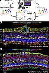
We generated a comparative spatial proteomic atlas of the human and mouse retina using a highly multiplexed immunohistochemistry technique called iterative bleaching extends multiplexity (IBEX). We refined the IBEX workflow by integrating an antibody dissociation option alongside chemical bleaching. This dual strategy enabled removal of the entire antibody complex, permitting the flexible use of antibodies from the same host species across iterative cycles. We coupled this workflow with super-resolution imaging via deconvolution and applied it to the retina of healthy humans and WT mice and the Crb1rd8 mouse model. We successfully imaged over 25 protein markers on human and mouse tissue sections, generating spatial atlases of the major retinal cell populations. Cross-species protein expression was compared to scRNA-seq datasets to identify protein and transcript disparities. Super-resolution IBEX delineated the ultrastructural features of the outer limiting membrane (OLM), identifying CD44 as a core structural component tightly colocalized with a highly organized F-actin belt within Müller glial endfeet. Using the Crb1rd8 mouse model, disruption of this complex was spatially associated with rosette formation and OLM structural failure. In summary, spatial proteomic atlases of the human and mouse retina were used to reveal insights into the arrangement of major retinal cell populations and OLM structure.
Authors
Yuxuan Meng, Jakub Kubiak, Zuzanna Dzieniak, Lorna Fowler, Rose Avient, Jason Hopley, Linyulong Li, Chaoran Li, Yuan Tian, Bruno Charbit, Colin J. Chu
Abstract
Systemic inflammation is now recognized as a key contributor to epilepsy pathophysiology, yet the role of innate immune cells, particularly neutrophils, remains poorly defined in epilepsy. While preclinical studies in rodent models have implicated neutrophils in seizure activity, their phenotype in human epilepsy has not been thoroughly investigated. In this study, we aimed to characterize systemic inflammatory profiles and neutrophil-associated immune signatures in the blood of patients with drug-resistant epilepsy, compared to healthy controls. We identified a systemic low-grade inflammatory profile in patients, characterized by elevated neutrophil-to-lymphocyte ratio, C-reactive protein, pro-inflammatory cytokines (IL-6, CXCL8/IL-8, TNF-α), and activated neutrophils (CXCR4+CD62Llow). Neutrophil phenotyping revealed two distinct immune profiles. Patients with longer disease duration exhibited a more immature systemic signature, characterized by immature neutrophils (CD15⁺CD10⁻), resting neutrophils (CXCR4⁺CD62L⁺), and elevated IL-6 levels. In contrast, patients with higher seizure frequency displayed a more inflammatory profile, marked by increased IL-12 and activated (CXCR4+CD62Llow) and hyperactivated (CXCR4highCD62Llow) neutrophil subsets. Moreover, elevated pre-surgical levels of inflammatory profile TNF-α, IL-6, and hyperactivated CXCR4high CD62Llow neutrophils were associated with seizure recurrence one year after surgery. This pioneering study highlights the heterogeneity of peripheral immune responses in drug-resistant epilepsy and identifies neutrophil-related signatures as promising prognostic biomarkers in this context.
Authors
Coraly Simoës Da Gama, Aurelie Hanin, Gwen Goudard, Veronique Masson, Aurore Besnard, Karim Dorgham, Guy Gorochov, Guillaume Dorothee, Valerio Frazzini, Vincent Navarro, Mélanie Morin-Brureau
Abstract
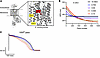
Short QT syndrome is a heritable arrhythmia disorder linked to sudden cardiac death. We recently identified that individuals with alternating hemiplegia of childhood (AHC), a rare neurodevelopmental disorder, can exhibit shortened corrected QT intervals and elevated risk for ventricular fibrillation. This is especially true for patients with AHC heterozygous for the recurrent ATP1A3-D801N variant, though the underlying cardiac mechanism remains unclear. We hypothesized that the D801N missense impairs Na+/K+-ATPase function, causing Ca2+ overload, shortened action potential duration (APD), and arrhythmias. Using in silico modeling and patient-derived induced pluripotent stem cell cardiomyocytes (iPSC-CMsD801N), we observed shorter APD, elevated intracellular and sarcoplasmic reticulum Ca2+ levels, and delayed afterdepolarizations (DADs) compared with WT. Additionally, increased Ca²+ influx via the Na+/Ca2+ exchanger (NCX1) during depolarization was observed in iPSC-CMsD801N. Simulations and in vitro experiments suggest that reduced ATPase function accelerated inactivation of L-type Ca2+ channels. Pharmacologic inhibition of NCX1 with ORM-10103 normalized APD and reduced DADs. These findings support a Ca2+-mediated mechanism for arrhythmogenesis in ATP1A3-D801N carriers and identify NCX1 as a potential therapeutic target.
Authors
Minu-Tshyeto K. Bidzimou, Padmapriya Muralidharan, Zhushan Zhang, Danyal Raza, Daniel Needs, Bo Sun, Robin M. Perelli, Mary E. Moya-Mendez, P.K. Rakesh Manivannan, Arsen S. Hunanyan, Abbigail Helfer, Christine Q. Simmons, Alfred L. George Jr., Donald M. Bers, Nenad Bursac, Mohamad A. Mikati, Andrew P. Landstrom

Abstract
Current treatment protocols for most types of cancers require chemotherapeutic agents that are associated with significant side effects, including chemotherapy-induced peripheral neuropathy (CIPN). Currently, there are no effective CIPN prevention strategies, and current treatment approaches remain limited. The enzyme purine nucleoside phosphorylase (PNPase) actively modulates both oxidative injury and cellular damage. Here, we tested the hypothesis that the signs and symptoms of CIPN are due to a chemotherapy-induced dysregulation of the purine metabolome. We assessed the effect of PNPase inhibition on paclitaxel-induced (PAC-induced) CIPN. Female adult Sprague-Dawley rats were treated with PAC and randomized to oral treatment with either the PNPase inhibitor 8-aminoguanine (8-AG) or its vehicle. Some rats were injected with shRNA against PNPase prior to PAC injections. PAC-treated rats exhibited multiple abnormalities: mechanical allodynia and changes in damaging purines, intraepidermal nerve fiber (IENF) density, and signaling cascades involved in mitochondrial disruption and axonal damage. Inhibition of PNPase improved behavioral function (mechanical allodynia), rescued the loss/damage of IENF, and normalized markers for mitochondrial dysfunction and nerve damage. These findings support the hypothesis that inhibition of PNPase prevented (and potentially reversed) CIPN through several mechanisms that included a reduction in neuronal damage and development of mechanical allodynia.
Authors
Lori A. Birder, Amanda Wolf-Johnston, Jonathan Franks, Mara L.G. Sullivan, Simon C. Watkins, Anthony J. Kanai, Vladimir B. Ritov, Edwin K. Jackson
Abstract
Chronic neuropathic pain is frequently comorbid with anxiety disorders, yet the neural circuits underlying this interaction remain poorly defined. The parafascicular nucleus of the thalamus (PF) integrates nociceptive and affective signals, but its specific regulatory mechanisms in pain-anxiety comorbidity are not well known. Using spared nerve injury (SNI) model mice, we combined viral neural tracing, chemogenetics, pharmacology, and electrophysiology to dissect the locus coeruleus (LC)-PF neural pathway. Viral tracing revealed monosynaptic projections from norepinephrinergic (NEergic) neurons in the dorsal LC to calcium/calmodulin dependent protein kinase IIα (CaMKIIα)- immunopositive neurons within the PF. Chemogenetic inhibition/activation of this pathway were performed in naïve and SNI mice, alongside intra-PF microinjection of the alpha-2 adrenergic receptor (ADRA2) antagonist yohimbine. Behavioral tests assessed mechanical/thermal hypersensitivity and anxiety-like behaviors. Results showed that 92.1% of PF-projecting LC neurons were NEergic, with 70.1% localized dorsally. Chemogenetic inhibition of LCNE-PFCaMKIIα neural pathway significantly alleviated both acute-phase mechanical hypersensitivity (< 7 days post-surgery) and chronic-phase anxiety-like behaviors in SNI mice, while activation of this pathway induced pain sensitization and anxiety-like behaviors in naïve mice. Intra-PF yohimbine reversed SNI-induced allodynia and anxiety-like behaviors. Electrophysiology confirmed yohimbine increased PF neuronal intrinsic excitability. These results suggest that the LCNE-PFCaMKIIα neural pathway promotes neuropathic pain and comorbid anxiety via ADRA2-mediated suppression of PF neuronal activity. Targeted inhibition of this circuit may represent a therapeutic strategy for pain-related affective disorders.
Authors
Zhong-Yi Liu, Fei Li, Li-Ming Liu, Yao-Hua Liu, Jia Li, Zi-Ang Li, Jin Cheng, Tian-Yu Zhao, Hui-Min Tian, Dong-Ning Li, Sha-Sha Tao, Hui Li, Fen-Sheng Huang, Yun-Qing Li
No posts were found with this tag.


