
Immunology
Abstract
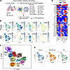
Idiopathic pulmonary fibrosis (IPF) is a fatal interstitial lung disease characterized by progressive scarring and respiratory failure. While T cells are elevated in IPF lungs, their contributions to fibrosis beyond inflammation remain poorly understood. Here, we performed multiplex imaging and single-cell RNA and protein profiling on about 90,000 CD3+ T cells from control and fibrotic lungs, revealing 11 distinct subsets of CD4+ and CD8+ T cells, including a rare CD56+ regulatory T cell. In addition to increased T cell numbers in severely fibrotic lungs compared with non-diseased controls, we observed CD4+ and CD8+ T cells localized near epithelial cells and in niches of abnormal epithelium. CXCR4/MIF signaling emerged as a central axis mediating T cell–epithelial interactions, while epidermal growth factor receptor (EGFR) and TGF-β pathways dominated in multiple T cell subsets. Our findings support the concept that T cells in IPF adopt nonclassical activation patterns that are driven by epithelial interactions within the fibrotic microenvironment. These studies provide a foundation for exploring alternative therapeutic strategies in IPF lungs by modulating T cell behavior and communication networks.
Authors
Ana P.M. Serezani, Julia M.R. Bazzano, Bruno D. Pascoalino, Ludmilla da Silva, Abigail J. Dietrich, Chase J. Taylor, Taylor Sherrill, Annika Vannan, Carla L. Calvi, Paula I. Gonzalez-Ericsson, Erin M. Wilfong, Matthew Bacchetta, Ciara M. Shaver, Lorraine B. Ware, Margaret L. Salisbury, Luc Van Kaer, Nicholas E. Banovich, Jonathan A. Kropski, Timothy S. Blackwell
Abstract
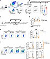
The cholinergic antiinflammatory pathway attenuates lung inflammation via the α7 nicotinic acetylcholine receptor (α7 nAChR) on immune cells. However, the role of α7 nAChR on lung megakaryocytes (Mks) in allergic airway inflammation remains unknown. In this study, allergen-challenged mouse models were used with conditional Mk-specific Chrna7 knockout, pharmacological activation (GTS-21), and Mk reconstitution. IL-33 expression and p38 MAPK signaling were assessed. We found that allergen challenge upregulated α7 nAChR specifically in lung Mks. Mk-specific Chrna7 deletion significantly alleviated allergic airway inflammation, whereas GTS-21 exacerbated inflammation via an Mk-dependent mechanism. Reconstitution with α7 nAChR+ Mks restored airway inflammatory responses. Mechanistically, α7 nAChR activation promoted Mk IL-33 synthesis and secretion through p38 MAPK signaling. Taken together, our results show that α7 nAChR on lung Mks plays a proinflammatory role in allergic airway inflammation, challenging its classical antiinflammatory paradigm and revealing pathogenic mechanisms.
Authors
Hang Wu, Rujia Tao, Shitao Xie, Yao Zhou, Jin-Fu Xu, Zhenwei Xia, Xiao Su

Abstract
Gain-of-function (GOF) variants in STAT3 cause a complex disorder characterized by early-onset autoimmunity, lymphoproliferation, recurrent infections, and immune dysregulation. In both primary human and mouse models of STAT3 GOF, CD8+ T cells have been implicated as pathogenic drivers of autoimmunity, though the exact mechanisms remain poorly understood. Here, we found that in patients with STAT3 GOF, CD8+ T cells exist in an activated state. Functional assessment revealed that naive CD8+ T cells have an increased capacity for IFN-γ and TNF-α production, with type I and type II IFN transcriptional signatures. Evaluation of immunoregulatory pathways revealed dysregulation of the purinergic signaling axis in CD8+ T cells: CD39 was increased, whereas downstream purinergic family members, CD73 and the adenosine receptor A2AR, were downregulated, impairing the potential to produce or sense immunosuppressive adenosine. Evaluation of the impact of precision therapy, in the form of JAK inhibition, at a cellular and functional level revealed partial normalization of CD8+ T cell dysregulation in patients, including aberrant cytokine production. Our study suggests that a dysregulated purinergic signaling axis plays a key role in CD8+ T cell dysregulation in STAT3 GOF and may have implications for other rare monogenic immune disorders and common inflammatory disorders.
Authors
Jose S. Campos Duran, Montana S. Knight, Samir U. Sayed, Megan C. Dalalo, Andrea A. Mauracher, Peyton Conrey, Aaron B. Schultz, Ceire A. Hay, Robert B. Lindell, Ilona Neale, Kyle Yeakle, Eric D. Abrams, Erica G. Schmitt, Martin A. Thelin, Christian A. Howard, Sara Bluestein, Christine M. Seroogy, Tamara C. Pozos, Akaluck Thatayatikom, Ingrid S. Lundgren, Amelie Gauthier, Scott W. Canna, Helen C. Su, Michael D. Keller, Ottavia M. Delmonte, Lisa R. Forbes Satter, Steven M. Holland, Jenna R.E. Bergerson, Jennifer W. Leiding, Neil Romberg, Will Bailis, Christopher A. Hunter, Alexandra F. Freeman, Alejandro V. Villarino, Mark S. Anderson, Megan A. Cooper, Tiphanie P. Vogel, Sarah E. Henrickson
Abstract
Clinical immunity to malaria develops after repeated malaria episodes. In this process, the inflammatory response is modulated to respond less vigorously upon reinfection. Monocytes are a major source of pro-inflammatory mediators during blood-stage infection and are known to adapt to repeated pathogen exposure. Here, we investigated the impact of previous malaria exposure on monocytes during blood-stage malaria by comparing the response in previously exposed and primary infected individuals. We observed reduced levels of several proinflammatory chemokines in previously exposed individuals, linked to changes in monocytes. Similarly, BAFF levels were lower in these individuals and associated with modulation of monocyte and dendritic cells. This affected the BAFF-BAFF-R axis, crucial for B cell responses, correlating with increasing parasite-specific antibody levels. Collectively, we present insights into how previous malaria exposure shapes monocyte responses during acute malaria and how these in turn correlate with modulation of the B cell compartment and humoral immune response.
Authors
Maximilian Julius Lautenbach, Pengjun Xi, Linn Kleberg, Alan-Dine Courey-Ghaouzi, Maia Serene Gower, Carolina Sousa Silva, Felicia Chammas, Anna Färnert, Christopher Sundling
Abstract
Autoimmune kidney diseases can cause glomerulonephritis and tubulointerstitial nephritis, which if unresolved, lead to progressive glomerulosclerosis and tubulointerstitial fibrosis. The IL-1 receptor (IL-1R1) is known to have divergent and cell-specific effects in kidney injury. We hypothesized that IL-1R1 would dampen pro-inflammatory activation of myeloid cells such that deletion of myeloid cell IL-1R1 would exacerbate autoimmune nephritis. Mice with myeloid cell-specific deletion of IL-1R1 (LysMCre(+) / Il1r1fl/fl - MKO) and littermate controls (LysMCre(-) / Il1r1fl/fl - MWT) were subjected to nephrotoxic serum (NTS) nephritis. MKO mice demonstrated worsened glomerular and tubular injury as indicated by increased albuminuria, glomerular injury scores, and kidney mRNA levels of kidney injury molecule (KIM)-1 (Havcr1) and neutrophil gelatinase-associated lipocalin (NGAL/Lcn2). We further found that myeloid IL-1R1 deficiency resulted in increased myeloid cell ER stress and expression of the heterodimeric cytokine Ebi3/Il27a (IL-27). IL-27 then induced increased type I IFN expression by kidney endothelial cells. In turn, anti-IL-27 limited type I IFN expression in endothelial cells and NTS nephritis, and anti-IFNAR1 therapy ameliorated glomerular and tubular injury in MKO mice. Thus, we demonstrated a myeloid cell-endothelial cell immunoregulatory axis whereby myeloid IL-1R1 activity constrained endothelial type I IFN generation to limit chronic kidney damage.
Authors
Yanting Chen, Yu Li, Jiafa Ren, Chia-Chun Wu, Xiaohan Lu, Achintya Inumarty, Steven D. Crowley, Jamie R. Privratsky
Abstract
Identifying and monitoring autoreactive T cells that drive beta cell destruction remains a major obstacle to developing effective immunotherapies for type 1 diabetes (T1D). These cells are extremely rare in peripheral blood and cannot be accessed directly from the pancreas. We used intradermal injection of Glutamic Acid Decarboxylase (GAD)-Alum to recruit GAD-specific T cells to accessible sites in the skin and skin-draining lymph nodes (LNs), sampled by skin suction blisters and ultrasound-guided LN aspiration. Peripheral blood samples obtained before GAD injection were restimulated with GAD in vitro to detect reactive CD4+ T cells. Single-cell RNA sequencing (scRNAseq) followed by re-expression of selected T cell receptors (TCRs) confirmed antigen specificity. Up to 70% of T cells at the skin injection site were clonally-expanded and 4 of 14 (28%) re-expressed TCRs were GAD-reactive. In LNs 1 of 14 (4%) clonally-expanded TCRs was GAD-reactive, representing ~0.08% of all T-cells. GAD-reactive cells across compartments displayed Th1 and Th17-associated transcription signatures. These results demonstrate the intradermal autoantigen challenge and scRNAseq, enable direct identification and molecular profiling of autoreactive T cells in vivo. This minimally invasive approach provides a powerful platform for tracking antigen-specific T cells to monitor disease activity and evaluate immune interventions in T1D.
Authors
Stephanie J. Hanna, Emma J.S. Robinson, Terri C. Thayer, Maki Nakayama, Laurie Landry, Robert Andrews, Garry Dolton, Joanne Davies, Evangelia Williams, James A. Pearson, Andrew K. Sewell, Parth Narendran, David Wraith, Alexandra Howell, Philippa Young, Mary Hart, Anton Lindqvist, F. Susan Wong, Tim I.M. Tree, Colin M. Dayan, Danijela Tatovic
Abstract
Allergic contact dermatitis (ACD), a recurrent inflammatory skin disorder, affects 21% of humans and is the second leading cause of occupational diseases in USA. ACD is initiated by the innate immune response to skin-contact sensitizers potentiated by the neuropeptide substance P (SP). Skin sensitizers stimulate SP-secreting sensory nerves and trigger proinflammatory functions of keratinocytes expressing the neurokinin 1 receptor (NK1R). Nevertheless, the neuroimmune regulation of hapten-initiated skin inflammation, remains incompletely elucidated. Using K14Cre/+NK1RKO mice skin-sensitized with 2,4-dinitrochlorobenzene (DNCB), we demonstrate that NK1R deletion exclusively in keratinocytes prevents hapten-initiated skin inflammation, impairs the mobilization of conventional dendritic cells (cDC) to draining lymph nodes (dLN) and blocks the elicitation of the contact hypersensitivity reaction (CHS) to the same extent observed in global Tac1KO (without SP) and NK1RKO mice. The DNCB effects were restored by skin co-administration of IL-1β and TNF-α. SP-NK1R signaling of mouse and human keratinocytes increased transcripts encoding proteins of the NLRP3 inflammasome. Although, DNCB and SP induced pro-IL-1β synthesis, only SP triggered intracellular Ca2+ increase, NFATc1 nuclear translocation and TNF-α synthesis, a cytokine mediating systemic inflammation in ACD. Our data identifying SP-NK1R-signaling of keratinocytes as a key mechanism for ACD provide relevant insight for therapies targeting skin neuroimmune interactions.
Authors
Sumeet Manandhar, Mohna Bandyopadhyay, Olga Tkacheva, William Shufesky, Greg Gibson, Simon C. Watkins, Adrian Morelli, Adriana T. Larregina
Abstract
DNA ligase IV (LIG4) is essential for DNA double-strand break (DSB) repair. Hypomorphic LIG4 variants cause LIG4 syndrome, characterized by growth disturbance, increased radiosensitivity, predisposition to malignancies, adaptive immunodeficiency and inflammatory conditions. Most of these manifestations are recapitulated in hypomorphic LIG4 mutant mice. However, no model mice with defective DSB repair have consistently exhibited inflammation. Here, we have generated mutant mice carrying the LIG4 missense variant, p.W447C, found in a patient with LIG4 syndrome. Lig4W447C/W447C mice showed functional defects of LIGIV and manifested growth retardation, increased radiosensitivity, and life-threatening intestinal inflammation under severe adaptive immunodeficiency. The inflammation was dependent on lymphocytes and characterized by marked infiltration of Th1 cells and macrophages, along with elevated expression of IFN-γ-inducible genes. When Ifng was deleted, Th2 and Th17 instead of Th1 cells drove the inflammation. Single-cell RNA-seq analyses with TCR repertoire revealed that T cells from Lig4W447C/W447C mice preferentially used proximal Vα and Jα segments in V regions of TCRα chains and exhibited expansion of several clonotypes, a substantial portion of which were CD4 T cells expressing IFN-γ. Thus, our hypomorphic Lig4 mutant mice represent a unique model for studying Th1-skewed intestinal inflammation under severe adaptive immunodeficiency.
Authors
Yusuke Yamashita, Hideki Kosako, Takashi Kato, Izumi Sasaki, Sadahiro Iwabuchi, Yuri Fukuda-Ohta, Tadashi Okamura, Misato Tane, Shotaro Tabata, Kazutaka Nakashima, Ken Tanaka, Kazunori Shiraishi, Yuki Uchihara, Daisuke Okuzaki, Kyoichi Isono, Atsushi Shibata, Tsunehiro Mizushima, Hiroaki Hemmi, Nobuo Kanazawa, Seiji Kodama, Hiroaki Miyoshi, Koichi Ohshima, Shinichi Hashimoto, Yoshio Fujitani, Takashi Sonoki, Shinobu Tamura, Tsuneyasu Kaisho
Abstract
Rationale: Persistent monocyte activation contributes to HIV-associated neurocognitive disorders (HAND), yet biomarkers that predict neurocognitive impairment before and after antiretroviral therapy (ART) remain incompletely defined. Objectives: We evaluated monocyte subsets and activation markers in participants from the SEARCH007 cohort prior to ART initiation and at 6 and 12 months following treatment. Methods and Results: Increased frequencies of CD14+CD16+ monocytes and elevated CD163 expression were associated with worsening neurocognitive performance and HAND severity. Plasma soluble CD163 levels increased with neurocognitive impairment and correlated with plasma HIV RNA levels, while CCR2 expression was associated with NPZ Global scores. Notably, CD169 expression was elevated across all monocyte subsets and demonstrated a stepwise increase with worsening neurocognitive impairment. Although ART reduced overall monocyte activation, elevated CD169 expression persisted in some individuals despite virologic suppression. Bayesian kernel machine regression and random forest analyses identified CD169 expression as one of the strongest predictors of cognitive impairment, surpassing plasma viral load, CD4+ T-cell count, and several established monocyte activation markers. Conclusions: These findings identify monocyte CD169 expression as a biomarker of neurocognitive dysfunction before and during the first year of ART and support further investigation of its role in HAND pathogenesis.
Authors
Hai Duc Nguyen, Andrew K. Ding-Su, Caroline Soulas, Tricia H. Burdo, Patrick Autissier, Pasiri Sithinamsuwan, Nitiya Chomchey, Jintanat Ananworanich, Victor Valcour, Silvia Ratto-Kim, Woong-Ki Kim, Kenneth C. Williams

Abstract
Authors
Xiao Yang, Sameen Fatima, Salvador Sampere-Birlanga, Akshay Ware, Mariana Shumliakivska, Lukas Zanders, Srisurekha Radhakrishnan, Guillermo Luxán, David John, Stefan Günther, Silvia Mas-Peiro, Stefanie Dimmeler, Andreas M. Zeiher, Wesley T. Abplanalp
No posts were found with this tag.


