
Research ArticleDevelopmentGeneticsNephrology
Open Access |  10.1172/jci.insight.203999
Constitutive YAP activation in distal nephron segments disrupts epithelial identity and nephron patterning
Zeinab Dehghani-Ghobadi,1,2 Eunah Chung,1,2 Mohammed Sayed,3 Christopher Ahn,3 Hyojin Alex Choi,1,2 Annissa Aamoum,1,2 Benjamin R. Thomson,1,4,5 Yueh-Chiang Hu,6,7 Hee-Woong Lim,3,7 and Joo-Seop Park1,2
1Feinberg Cardiovascular & Renal Research Institute and
2Division of Nephrology and Hypertension, Department of Medicine, Northwestern University Feinberg School of Medicine, Chicago, Illinois, USA.
3Division of Biomedical Informatics, Cincinnati Children’s Hospital Medical Center, Cincinnati, Ohio, USA.
4Department of Ophthalmology, Northwestern University Feinberg School of Medicine, Chicago, Illinois, USA.
5Department of Biomedical Engineering, Northwestern University, Evanston, Illinois, USA.
6Division of Developmental Biology, Cincinnati Children’s Hospital Medical Center, Cincinnati, Ohio, USA.
7Department of Pediatrics, University of Cincinnati College of Medicine, Cincinnati, Ohio, USA.
Address correspondence to: Joo-Seop Park, Division of Nephrology and Hypertension, Feinberg Cardiovascular & Renal Research Institute, Northwestern University Feinberg School of Medicine, 303 E. Superior St., SQBRC 8-406, Chicago, Illinois 60611, USA. Email: jooseop.park@northwestern.edu.
Find articles by Dehghani-Ghobadi, Z. in: PubMed | Google Scholar
1Feinberg Cardiovascular & Renal Research Institute and
2Division of Nephrology and Hypertension, Department of Medicine, Northwestern University Feinberg School of Medicine, Chicago, Illinois, USA.
3Division of Biomedical Informatics, Cincinnati Children’s Hospital Medical Center, Cincinnati, Ohio, USA.
4Department of Ophthalmology, Northwestern University Feinberg School of Medicine, Chicago, Illinois, USA.
5Department of Biomedical Engineering, Northwestern University, Evanston, Illinois, USA.
6Division of Developmental Biology, Cincinnati Children’s Hospital Medical Center, Cincinnati, Ohio, USA.
7Department of Pediatrics, University of Cincinnati College of Medicine, Cincinnati, Ohio, USA.
Address correspondence to: Joo-Seop Park, Division of Nephrology and Hypertension, Feinberg Cardiovascular & Renal Research Institute, Northwestern University Feinberg School of Medicine, 303 E. Superior St., SQBRC 8-406, Chicago, Illinois 60611, USA. Email: jooseop.park@northwestern.edu.
Find articles by Chung, E. in: PubMed | Google Scholar
1Feinberg Cardiovascular & Renal Research Institute and
2Division of Nephrology and Hypertension, Department of Medicine, Northwestern University Feinberg School of Medicine, Chicago, Illinois, USA.
3Division of Biomedical Informatics, Cincinnati Children’s Hospital Medical Center, Cincinnati, Ohio, USA.
4Department of Ophthalmology, Northwestern University Feinberg School of Medicine, Chicago, Illinois, USA.
5Department of Biomedical Engineering, Northwestern University, Evanston, Illinois, USA.
6Division of Developmental Biology, Cincinnati Children’s Hospital Medical Center, Cincinnati, Ohio, USA.
7Department of Pediatrics, University of Cincinnati College of Medicine, Cincinnati, Ohio, USA.
Address correspondence to: Joo-Seop Park, Division of Nephrology and Hypertension, Feinberg Cardiovascular & Renal Research Institute, Northwestern University Feinberg School of Medicine, 303 E. Superior St., SQBRC 8-406, Chicago, Illinois 60611, USA. Email: jooseop.park@northwestern.edu.
Find articles by Sayed, M. in: PubMed | Google Scholar
1Feinberg Cardiovascular & Renal Research Institute and
2Division of Nephrology and Hypertension, Department of Medicine, Northwestern University Feinberg School of Medicine, Chicago, Illinois, USA.
3Division of Biomedical Informatics, Cincinnati Children’s Hospital Medical Center, Cincinnati, Ohio, USA.
4Department of Ophthalmology, Northwestern University Feinberg School of Medicine, Chicago, Illinois, USA.
5Department of Biomedical Engineering, Northwestern University, Evanston, Illinois, USA.
6Division of Developmental Biology, Cincinnati Children’s Hospital Medical Center, Cincinnati, Ohio, USA.
7Department of Pediatrics, University of Cincinnati College of Medicine, Cincinnati, Ohio, USA.
Address correspondence to: Joo-Seop Park, Division of Nephrology and Hypertension, Feinberg Cardiovascular & Renal Research Institute, Northwestern University Feinberg School of Medicine, 303 E. Superior St., SQBRC 8-406, Chicago, Illinois 60611, USA. Email: jooseop.park@northwestern.edu.
Find articles by Ahn, C. in: PubMed | Google Scholar
1Feinberg Cardiovascular & Renal Research Institute and
2Division of Nephrology and Hypertension, Department of Medicine, Northwestern University Feinberg School of Medicine, Chicago, Illinois, USA.
3Division of Biomedical Informatics, Cincinnati Children’s Hospital Medical Center, Cincinnati, Ohio, USA.
4Department of Ophthalmology, Northwestern University Feinberg School of Medicine, Chicago, Illinois, USA.
5Department of Biomedical Engineering, Northwestern University, Evanston, Illinois, USA.
6Division of Developmental Biology, Cincinnati Children’s Hospital Medical Center, Cincinnati, Ohio, USA.
7Department of Pediatrics, University of Cincinnati College of Medicine, Cincinnati, Ohio, USA.
Address correspondence to: Joo-Seop Park, Division of Nephrology and Hypertension, Feinberg Cardiovascular & Renal Research Institute, Northwestern University Feinberg School of Medicine, 303 E. Superior St., SQBRC 8-406, Chicago, Illinois 60611, USA. Email: jooseop.park@northwestern.edu.
Find articles by Choi, H. in: PubMed | Google Scholar
1Feinberg Cardiovascular & Renal Research Institute and
2Division of Nephrology and Hypertension, Department of Medicine, Northwestern University Feinberg School of Medicine, Chicago, Illinois, USA.
3Division of Biomedical Informatics, Cincinnati Children’s Hospital Medical Center, Cincinnati, Ohio, USA.
4Department of Ophthalmology, Northwestern University Feinberg School of Medicine, Chicago, Illinois, USA.
5Department of Biomedical Engineering, Northwestern University, Evanston, Illinois, USA.
6Division of Developmental Biology, Cincinnati Children’s Hospital Medical Center, Cincinnati, Ohio, USA.
7Department of Pediatrics, University of Cincinnati College of Medicine, Cincinnati, Ohio, USA.
Address correspondence to: Joo-Seop Park, Division of Nephrology and Hypertension, Feinberg Cardiovascular & Renal Research Institute, Northwestern University Feinberg School of Medicine, 303 E. Superior St., SQBRC 8-406, Chicago, Illinois 60611, USA. Email: jooseop.park@northwestern.edu.
Find articles by Aamoum, A. in: PubMed | Google Scholar
1Feinberg Cardiovascular & Renal Research Institute and
2Division of Nephrology and Hypertension, Department of Medicine, Northwestern University Feinberg School of Medicine, Chicago, Illinois, USA.
3Division of Biomedical Informatics, Cincinnati Children’s Hospital Medical Center, Cincinnati, Ohio, USA.
4Department of Ophthalmology, Northwestern University Feinberg School of Medicine, Chicago, Illinois, USA.
5Department of Biomedical Engineering, Northwestern University, Evanston, Illinois, USA.
6Division of Developmental Biology, Cincinnati Children’s Hospital Medical Center, Cincinnati, Ohio, USA.
7Department of Pediatrics, University of Cincinnati College of Medicine, Cincinnati, Ohio, USA.
Address correspondence to: Joo-Seop Park, Division of Nephrology and Hypertension, Feinberg Cardiovascular & Renal Research Institute, Northwestern University Feinberg School of Medicine, 303 E. Superior St., SQBRC 8-406, Chicago, Illinois 60611, USA. Email: jooseop.park@northwestern.edu.
Find articles by
Thomson, B.
in:
PubMed
|
Google Scholar
|

1Feinberg Cardiovascular & Renal Research Institute and
2Division of Nephrology and Hypertension, Department of Medicine, Northwestern University Feinberg School of Medicine, Chicago, Illinois, USA.
3Division of Biomedical Informatics, Cincinnati Children’s Hospital Medical Center, Cincinnati, Ohio, USA.
4Department of Ophthalmology, Northwestern University Feinberg School of Medicine, Chicago, Illinois, USA.
5Department of Biomedical Engineering, Northwestern University, Evanston, Illinois, USA.
6Division of Developmental Biology, Cincinnati Children’s Hospital Medical Center, Cincinnati, Ohio, USA.
7Department of Pediatrics, University of Cincinnati College of Medicine, Cincinnati, Ohio, USA.
Address correspondence to: Joo-Seop Park, Division of Nephrology and Hypertension, Feinberg Cardiovascular & Renal Research Institute, Northwestern University Feinberg School of Medicine, 303 E. Superior St., SQBRC 8-406, Chicago, Illinois 60611, USA. Email: jooseop.park@northwestern.edu.
Find articles by Hu, Y. in: PubMed | Google Scholar
1Feinberg Cardiovascular & Renal Research Institute and
2Division of Nephrology and Hypertension, Department of Medicine, Northwestern University Feinberg School of Medicine, Chicago, Illinois, USA.
3Division of Biomedical Informatics, Cincinnati Children’s Hospital Medical Center, Cincinnati, Ohio, USA.
4Department of Ophthalmology, Northwestern University Feinberg School of Medicine, Chicago, Illinois, USA.
5Department of Biomedical Engineering, Northwestern University, Evanston, Illinois, USA.
6Division of Developmental Biology, Cincinnati Children’s Hospital Medical Center, Cincinnati, Ohio, USA.
7Department of Pediatrics, University of Cincinnati College of Medicine, Cincinnati, Ohio, USA.
Address correspondence to: Joo-Seop Park, Division of Nephrology and Hypertension, Feinberg Cardiovascular & Renal Research Institute, Northwestern University Feinberg School of Medicine, 303 E. Superior St., SQBRC 8-406, Chicago, Illinois 60611, USA. Email: jooseop.park@northwestern.edu.
Find articles by
Lim, H.
in:
PubMed
|
Google Scholar
|
1Feinberg Cardiovascular & Renal Research Institute and
2Division of Nephrology and Hypertension, Department of Medicine, Northwestern University Feinberg School of Medicine, Chicago, Illinois, USA.
3Division of Biomedical Informatics, Cincinnati Children’s Hospital Medical Center, Cincinnati, Ohio, USA.
4Department of Ophthalmology, Northwestern University Feinberg School of Medicine, Chicago, Illinois, USA.
5Department of Biomedical Engineering, Northwestern University, Evanston, Illinois, USA.
6Division of Developmental Biology, Cincinnati Children’s Hospital Medical Center, Cincinnati, Ohio, USA.
7Department of Pediatrics, University of Cincinnati College of Medicine, Cincinnati, Ohio, USA.
Address correspondence to: Joo-Seop Park, Division of Nephrology and Hypertension, Feinberg Cardiovascular & Renal Research Institute, Northwestern University Feinberg School of Medicine, 303 E. Superior St., SQBRC 8-406, Chicago, Illinois 60611, USA. Email: jooseop.park@northwestern.edu.
Find articles by
Park, J.
in:
PubMed
|
Google Scholar
|
Published June 30, 2026 - More info
JCI Insight. 2026;11(16):e203999. https://doi.org/10.1172/jci.insight.203999.
© 2026 Dehghani-Ghobadi et al. This work is licensed under the Creative Commons Attribution 4.0 International License. To view a copy of this license, visit http://creativecommons.org/licenses/by/4.0/.
Received: December 29, 2025; Accepted: June 24, 2026

-
Results
DCT cells give rise to CNT cells during nephron development. The DCT of the nephron can be targeted using the previously reported Slc12a3-IRES-CreERT2 allele (20). However, because nephrogenesis occurs in waves during development, DCT cells are generated at different times, making it impractical for a tamoxifen-inducible system to achieve complete and uniform targeting of all DCT cells. To overcome this limitation, we generated Slc12a3-IRES-Cre (hereafter Slc12a3Cre) using CRISPR/Cas9 genome editing to convert Slc12a3-IRES-CreERT2 into a constitutively active allele. We assessed the recombinase activity of this new Cre line by lineage tracing with a Rosa26-Sun1 reporter (21). As expected, all SLC12A3+ cells were positive for GFP, confirming that Slc12a3Cre efficiently targets the entire DCT segment, including DCT1, the transition zone, and DCT2 (Figure 1A, Supplemental Figure 1A, and Supplemental Figure 2; supplemental material available online with this article; https://doi.org/10.1172/jci.insight.203999DS1). Unexpectedly, we also observed GFP+ cells that lacked SLC12A3. Immunofluorescence analysis with distal nephron markers showed that these GFP+ SLC12A3– cells expressed S100G, suggesting that they were the CNT segment (Figure 1A, Supplemental Figure 1A, and Supplemental Figure 2). This observation suggests that CNT cells arise from the DCT lineage during development.
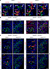 Figure 1
Constitutive activation of YAP in the DCT and CNT leads to increased proliferation and disrupts segmental identity. (A) Lineage tracing using the Rosa26-Sun1 reporter (nuclear membrane GFP) shows that Slc12a3Cre targets both DCT and CNT. SLC12A3 marks both DCT1 and DCT2; PVALB marks DCT1 only. S100G and CALB1 mark DCT2 and CNT. The transition zone (TZ) is marked by both DCT1 and DCT2 markers (see Supplemental Figure 2). Adjacent sections of the same kidney are shown. (B) In control kidneys, only a small subset of DCT and CNT cells (GFP+) are positive for Ki67, indicating low proliferative activity. In contrast, DCT and CNT cells with persistent YAP activation show strong Ki67 staining, consistent with increased proliferation. (C) GFP+ cells in the control kidney express markers characteristic of distal nephron segments, whereas GFP+ cells in the mutant kidney lack these markers, suggesting disrupted segmental identity. (A–C) Representative images from 3 independent experiments are shown. Stage, P6; scale bar: 100 μm.
Constitutive YAP activation in DCT and CNT cells increases proliferation and disrupts segmental identity. To establish the physiological context for YAP regulation in the distal nephron, we first examined YAP subcellular localization during early postnatal kidney development. Immunofluorescence analysis revealed that at P1, both proximal tubule and DCT/CNT cells exhibited nuclear YAP, whereas by P6, nuclear YAP was retained in proximal tubule cells but was largely absent from DCT/CNT cells (Supplemental Figure 3). These observations indicate that YAP activity is normally downregulated in distal tubule cells during postnatal maturation, defining a developmental stage at which sustained YAP activation would be non-native to distal nephron epithelia.
To test the consequences of preventing this developmental repression of YAP activity, we generated a Cre-dependent Yap gain-of-function (GOF) allele (Supplemental Figure 4). Upon Cre-mediated inversion, this allele expresses YAP5SA, a modified form in which 5 serine residues are replaced with alanine (22, 23). These substitutions prevent phosphorylation by Hippo kinases, thereby blocking YAP inactivation and rendering it constitutively active. Using CRISPR/Cas9 genome editing, we inserted the Cre-inducible Yap5SA cassette into the safe-harbor Col1a1 locus (hereafter Col1a1-Yap5SA; Supplemental Figure 4). Because constitutive YAP activation mimics Hippo pathway inhibition regardless of upstream inputs, this allele provides a powerful tool to assess the effects of sustained YAP activity in vivo.
To investigate the role of Hippo signaling in the distal nephron segments, we crossed Slc12a3Cre with Col1a1-Yap5SA to generate Yap GOF mutant kidneys, in which YAP was constitutively active in the DCT and CNT. A Rosa26-Sun1 reporter was included to mark cells that had experienced Cre-mediated recombination. Because the Yap GOF allele encodes a 3xFLAG-tagged YAP5SA protein, we first confirmed transgene expression within the targeted cell population by FLAG immunofluorescence. In mutant kidneys, all GFP+ distal nephron epithelial cells were FLAG positive, confirming robust Yap transgene expression in SLC12A3-lineage cells (Supplemental Figure 5).
At P6, mutant kidneys contained markedly increased numbers of GFP+ cells compared with controls, often arranged in multiple layers surrounding dilated lumens (Figure 1B; Supplemental Figure 1B). These GFP+ cells exhibited a significant increase in Ki67 staining, indicating hyperproliferation associated with constitutive YAP activation (Figure 1B; Supplemental Figure 1B). Immunofluorescence analysis using distal nephron markers revealed distinct segment-specific expression patterns in control kidneys but a near-complete loss of expression in mutant kidneys, consistent with disruption of distal nephron identity (Figure 1C and Supplemental Figure 1, C and D). S100G was still detected in mutant kidneys; however, the majority of S100G+ cells were GFP–, suggesting that they had not undergone sustained YAP activation (Figure 1C and Supplemental Figure 1D).
To determine the developmental onset of the phenotype, we next examined kidneys at E18.5 and P3. At E18.5, mutant kidneys exhibited early and heterogeneous defects in distal nephron identity, with subsets of GFP+ tubules lacking expression of distal segment markers (Supplemental Figure 6). By P3, loss of DCT and CNT marker expression became widespread among GFP+ cells, indicating a robust and uniform disruption of distal nephron segment identity (Supplemental Figure 7).
H&E staining further demonstrated pronounced tubular dilation and underdeveloped papillary architecture in mutant kidneys (Supplemental Figure 8A). Kaplan-Meier survival analysis revealed markedly increased neonatal mortality, with no mutant animals surviving beyond P7 (Supplemental Figure 8B).
Together, these data demonstrate that sustained YAP activation in distal nephron epithelial cells leads to early disruption of epithelial identity, followed by hyperproliferation, architectural disorganization, and early postnatal lethality.
Constitutive YAP activation disrupts polarity and junctional organization in DCT and CNT cells. Our observation that GFP+ cells in the mutant kidney often failed to remain as a monolayer prompted us to examine how constitutive YAP activation affects epithelial polarity and junctional organization. In control kidneys, GFP+ tubules showed LAMA1 at the basal side and PARD6B at the apical side (Figure 2, A and B). In contrast, GFP+ cells in mutant kidneys showed a marked loss of basal LAMA1 and disrupted apical distribution of PARD6B, indicating impaired polarity (Figure 2, A and B).
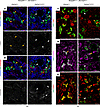 Figure 2
Constitutive activation of YAP in DCT and CNT impairs epithelial polarity and junctional organization. (A and B) In the control kidney, the basal and apical sides of GFP+ cells are marked by LAMA1 and PARD6B, respectively (orange arrowheads). In the mutant kidney, GFP+ cells (white arrowheads) lack basal LAMA1 and apical PARD6B. (C–E) In the control kidney, GFP+ cells express the tight junction marker TJP1 (ZO1) and the adherens junction markers, such as CTNNB1 and CDH1 (orange arrowheads). In mutant kidneys, GFP+ cells exhibit either a loss (white arrowheads) or mislocalization (orange arrowheads) of junctional markers, suggesting impaired epithelial integrity. (A–E) Representative images from 3 independent experiments are shown. Stage, P6; scale bar: 100 μm.
To determine when these polarity defects first arise during development, we examined distal nephron tubules at E18.5 and P3. At both stages, mutant kidneys exhibited heterogeneous polarity defects among GFP+ tubules, with some retaining polarized LAMA1 and PARD6B localization and others showing clear loss of polarity (Supplemental Figure 9A). By P3, a greater proportion of GFP+ tubules displayed polarity defects compared with E18.5, indicating a progressive loss of epithelial polarity during postnatal maturation (Supplemental Figure 9B).
Furthermore, in GFP+ cells in mutant kidneys, junctional proteins such as TJP1 (ZO-1), CTNNB1 (β-catenin), and CDH1 (E-cadherin) were frequently absent or mislocalized from their normal junctional positions (Figure 2, C–E, and Supplemental Figure 10), suggesting destabilization of cell-cell adhesion complexes. These alterations are consistent with YAP’s established role in maintaining epithelial architecture in other tissues (9, 24) and likely contribute to the loss of segment-specific identity in Yap GOF mutant kidneys.
Constitutive YAP activation impairs distal nephron identity and alters epithelial spatial organization. GATA3, a key transcription factor regulating the branching of the collecting duct during development (25, 26), is expressed in the DCT, CNT, and collecting duct of the renal epithelia (2). In Yap GOF mutant kidneys, GATA3 was largely absent in DCT and CNT cells marked by activation of the Rosa26-Sun1 reporter (Figure 3A and Supplemental Figure 11), indicating that sustained YAP activity disrupts segmental identity and differentiation in the distal nephron.
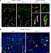 Figure 3
Constitutive YAP activation in DCT and CNT is associated with displacement of lineage-labeled cells beyond normal segmental boundaries. In both control and mutant kidneys, Slc12a3Cre activates GFP expression in DCT and CNT but not in the collecting duct (CD). (A) The left panel shows a low-magnification view of the kidney, with the regions outlined by dotted squares shown at higher magnification in the right panel. AQP2 marks CD; GATA3 marks DCT, CNT, CD, and mesangial cells. GFP+ cells in the control kidney are positive for GATA3, whereas those in the mutant kidney lack GATA3. In the mutant kidney, GFP+ cells are located within the lumen of AQP2+ GATA3+ CD cells. (B) SLC12A1 marks the ascending limb of the loop of Henle. In the mutant kidney, GFP+ cells are located within the lumen of SLC12A1+ cells (marked by yellow arrowheads). Representative images from 3 independent experiments are shown. Stage, P6; scale bar: 50 μm.
In addition to loss of segmental identity, GFP+ lineage-labeled distal epithelial cells were detected outside their expected segmental locations. Rosa26-Sun1 reporter–positive Yap GOF cells were observed within collecting duct structures marked by GATA3 and AQP2 (Figure 3A and Supplemental Figure 11). GFP+ cells were also identified within the thick ascending limb of the loop of Henle, as marked by SLC12A1 (Figure 3B), indicating altered spatial localization of distal nephron–derived cells relative to normal segmental boundaries.
The macula densa (MD) plays a central role in stimulating REN1 (renin) release from the juxtaglomerular apparatus (JGA) by sensing tubular NaCl delivery and producing paracrine mediators such as prostaglandin E2 (via COX-2) and nitric oxide (27). Because the MD represents a specialized subset of the thick ascending limb of the loop of Henle, it can be identified as SLC12A1+ cells positioned adjacent to the glomerulus (Supplemental Figure 12A). In Yap GOF mutant kidneys, GFP+ cells were observed within the lumen of the MD, indicating aberrant localization of distal nephron–derived cells within this specialized segment (Supplemental Figure 12A).
Consistent with disruption of MD/JGA signaling, REN1 signal was absent in the mutant kidneys (Supplemental Figure 12B). In parallel, RNA-Seq analysis revealed coordinated downregulation of multiple components of the renin-angiotensin system, including Ren1, Ace, Agtr1a, and Agtr2, in Yap GOF kidneys (Supplemental Table 1). Together, these findings indicate that constitutive YAP activation disrupts distal nephron organization and is associated with impaired MD-dependent endocrine signaling, providing a potential mechanistic link between distal epithelial disorganization and suppression of the MD/JGA axis.
To determine whether disruption of the MD/JGA axis is associated with impaired nephron flow, we performed an in vivo fluorescent dextran delivery assay at P3 (28). The MD/JGA axis is required for maintaining normal glomerular filtration by regulating REN1 release, intrarenal perfusion, and downstream tubular delivery. After i.v. administration of 10 kDa Texas red–conjugated dextran, control kidneys showed robust luminal dextran signal throughout nephron tubules, consistent with effective filtration and downstream delivery (Supplemental Figure 13). In contrast, mutant kidneys exhibited markedly reduced dextran signal, approaching background levels observed in noninjected controls (Supplemental Figure 13). These findings indicate that luminal delivery of filtrate is severely impaired in Yap GOF kidneys, consistent with a functional disruption of nephron flow along the tubular axis.
Kidney-wide transcriptomic responses to constitutive Yap activation in distal nephron segments. To evaluate the impact of constitutive YAP activation in DCT and CNT on kidney-wide transcriptional programs, we performed bulk RNA-Seq analysis on P3 kidneys from mutants and littermate controls (Supplemental Table 1). Our analysis identified 2,310 differentially expressed genes (DEGs), including 1,116 upregulated and 1,194 downregulated transcripts (fold change > 1.5, FDR < 0.05) (Supplemental Figure 14A). To determine the cellular origins of these DEGs, we mapped them onto our previously published single-cell RNA-Seq (scRNA-Seq) dataset of developing mouse kidneys (Supplemental Figure 14B). In the uniform manifold approximation and projection (UMAP), downregulated genes were enriched in clusters corresponding to the proximal tubule, thick ascending limb, and DCT (Supplemental Figure 14C). In contrast, upregulated genes were predominantly associated with clusters representing immune cell populations (Supplemental Figure 14D). Consistent with these assignments, Gene Ontology analysis showed that downregulated genes were strongly enriched for proximal tubule functions, including amino acid catabolism, fatty acid β-oxidation, and brush-border organization (Supplemental Figure 15A). In contrast, upregulated genes were enriched for inflammatory and immune-activation pathways, including TNF, MAPK, and NF-κB signaling, suggesting that YAP activation induces proinflammatory and stress-responsive transcriptional programs. Genes related to cell adhesion, cytoskeletal remodeling, and membrane-raft assembly were also elevated, consistent with altered epithelial architecture and cell-cell interactions in Yap GOF mutant kidneys (Supplemental Figure 15B).
Figure 4 highlights 3 groups of DEGs and their cell type–specific expression across the kidney: (a) downregulated DCT/CNT genes, (b) downregulated proximal tubule genes, and (c) upregulated injury-associated genes. Consistent with our findings of the loss of multiple DCT/CNT markers in the Yap GOF mutant kidneys (Figure 1C and Supplemental Figure 1C), bulk RNA-Seq confirmed their downregulation in the Yap GOF mutant kidneys (Figure 4).
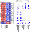 Figure 4
Differential gene expression analysis. Bulk RNA-Seq of whole kidneys at P3 reveals differentially expressed genes (DEGs) between mutant and control kidneys, as shown in the heatmap. In the mutant kidney, genes normally expressed in distal tubules and proximal tubules are downregulated, while injury-associated genes are upregulated. These expression changes occur regardless of sex. Color intensity represents the magnitude of expression change: blue indicates decreased expression, and red indicates increased expression. The corresponding dot plot displays cell type–specific expression of marker genes based on previously published scRNA-Seq data from mouse kidneys at E18.5 or P0 (GSE214024 and GSE275601). Dot color indicates the average expression level of each gene within a given cell type; dot size represents the percentage of cells expressing that gene.
We next validated the downregulation of proximal tubule genes in the Yap GOF mutant kidneys by immunofluorescence. Control kidneys showed strong Lotus tetragonolobus lectin (LTL) staining, reflecting the abundant glycoproteins in the brush border of mature proximal tubules (Figure 5). In contrast, mutant kidneys exhibited markedly reduced LTL labeling, indicating the absence of mature proximal tubules. Notably, HNF4A+ proximal tubule cells were still present in mutant kidneys, suggesting that proximal tubule specification still occurred. However, these cells failed to develop into mature proximal tubules. Consistent with impaired maturation, mature proximal tubule markers such as PCK1, FBP1, and SLC5A2 were nearly absent in mutant kidneys, and other proximal tubule markers were also reduced, though to a lesser degree.
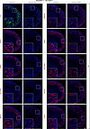 Figure 5
Constitutive YAP activation in DCT and CNT leads to the downregulation of proximal tubule genes. In the mutant kidney, LTL (Lotus tetragonolobus lectin) staining is greatly reduced compared with controls, indicating an absence of mature proximal tubules. Expression of proximal tubule markers, such as PCK1, SLC5A2, and FBP1, is almost absent in the mutant, along with the downregulation of additional proximal tubule genes. Inset panels, denoted by the white dotted squares, present higher-magnification views of the specified cortical regions outlined in the overview images. Representative images from 3 independent experiments are shown. Stage, P6; scale bar: 200 μm.
To determine the developmental onset of this proximal tubule maturation defect, we examined kidneys at E18.5 and P3 (Supplemental Figure 16). At E18.5, mutant kidneys showed preserved expression of proximal tubule markers (HNF4A, FBP1, and PCK1), accompanied by weaker LTL staining. By P3, mutant kidneys exhibited broader loss of proximal tubule marker expression and markedly diminished LTL signal, despite the continued presence of HNF4A+ cells. These findings indicate that proximal tubule specification is initiated but maturation progressively fails during late embryonic and early postnatal development in Yap GOF kidneys.
In addition to proximal tubule defects, the loop of Henle and collecting duct also exhibited structural abnormalities. Segment-specific marker analysis demonstrated impaired tubular segmentation, with markedly reduced expression of AQP1 (descending limb), SLC12A1 (thick ascending limb), and AQP2 (collecting duct principal cells) in mutant kidneys (Supplemental Figure 17). Taken together, these results suggest that YAP overactivation in the distal nephron segments blocks proximal tubule maturation, leading to a broad disruption of nephron patterning.
We found that injury-associated genes, including HAVCR1 (KIM1), SPP1, and LCN2 (NGAL), were markedly upregulated in mutant kidneys (Figure 4). These transcriptomic changes were corroborated by immunofluorescence staining, which showed increased HAVCR1 and SPP1 expression in proximal tubules, along with elevated KRT19 expression in distal nephron segments (Figure 6). Consistent with our observation that immune cell clusters in scRNA-Seq data show the enrichment of the genes upregulated in the Yap GOF mutant kidneys (Supplemental Figure 14D), the mutant kidneys exhibited enhanced macrophage infiltration around GFP+ Yap GOF mutant cells (Figure 6), suggesting that immune cell recruitment accompanies the injury response triggered by constitutive YAP activation. Together, these findings indicate that aberrant YAP signaling compromises nephron patterning and promotes inflammatory and cytoskeletal remodeling responses.
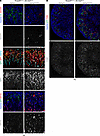 Figure 6
YAP activation in DCT and CNT cells induces tubular injury and macrophage infiltration. (A) In control kidneys, strong LTL labeling is observed with no detectable HAVCR1 signal. In contrast, mutant kidneys show reduced LTL labeling accompanied by HAVCR1 upregulation. Regions with weak HAVCR1 signal overlap with cells displaying weak LTL signal, whereas areas with strong HAVCR1 signal show no overlap with LTL. Two additional injury markers, SPP1 and KRT19, are upregulated in mutant kidneys. In control kidneys, SPP1 is largely expressed in HNF4A+ proximal tubules, whereas in mutant kidneys, SPP1 is upregulated in both HNF4A+ and HNF4A– tubules. In control kidneys, KRT19 signal is present in the papilla and absent in GFP+ cells, whereas in mutant kidneys, GFP+ cells show KRT19 upregulation. Stage, P6; scale bars: 100 μm. (B) Mutant kidneys show a pronounced increase in F4/80+ cells, a marker of mature tissue macrophages — reflecting enhanced macrophage infiltration and renal inflammation. Representative images from 3 independent experiments are shown. Stage, P6; scale bars: 200 μm.


