
Research ArticleImmunologyMuscle biology
Open Access |  10.1172/jci.insight.202541
CD8+ T cell activation occurs 24 hours after AAV administration and is driven by muscle promoter specificity
Lindsay Jeanpierre,1,2 Coralie Pecquet,1,2 Hanadi Saliba,1,2 Pauline Finard,1,2 Stéphane Terry,1,2,3 Gianni Tavella,1,2 Inès Guesmia,1,2 Sylvie Boutin,1,2 Bérangère Bertin,1,2 Sofia Benkhelifa-Ziyyat,4 Giuseppe Ronzitti,1,2 and David-Alexandre Gross1,2
1Généthon, Evry, France.
2Université Paris-Saclay, Université d’Évry, Inserm, Généthon, Integrare research unit UMR_S951, Evry, France.
3Research Department, Inovarion, Paris, France.
4Sorbonne Université, Inserm, Institut de Myologie, Centre de Recherche en Myologie, Paris, France.
Address correspondence to: David-Alexandre Gross, INSERM UMR_S951, Généthon, 1 bis rue de l’Internationale, 91000 Evry, France. Phone: 33.1.69.47.10.22; Email: david.gross@inserm.fr.
Find articles by Jeanpierre, L. in: PubMed | Google Scholar
1Généthon, Evry, France.
2Université Paris-Saclay, Université d’Évry, Inserm, Généthon, Integrare research unit UMR_S951, Evry, France.
3Research Department, Inovarion, Paris, France.
4Sorbonne Université, Inserm, Institut de Myologie, Centre de Recherche en Myologie, Paris, France.
Address correspondence to: David-Alexandre Gross, INSERM UMR_S951, Généthon, 1 bis rue de l’Internationale, 91000 Evry, France. Phone: 33.1.69.47.10.22; Email: david.gross@inserm.fr.
Find articles by Pecquet, C. in: PubMed | Google Scholar
1Généthon, Evry, France.
2Université Paris-Saclay, Université d’Évry, Inserm, Généthon, Integrare research unit UMR_S951, Evry, France.
3Research Department, Inovarion, Paris, France.
4Sorbonne Université, Inserm, Institut de Myologie, Centre de Recherche en Myologie, Paris, France.
Address correspondence to: David-Alexandre Gross, INSERM UMR_S951, Généthon, 1 bis rue de l’Internationale, 91000 Evry, France. Phone: 33.1.69.47.10.22; Email: david.gross@inserm.fr.
Find articles by Saliba, H. in: PubMed | Google Scholar
1Généthon, Evry, France.
2Université Paris-Saclay, Université d’Évry, Inserm, Généthon, Integrare research unit UMR_S951, Evry, France.
3Research Department, Inovarion, Paris, France.
4Sorbonne Université, Inserm, Institut de Myologie, Centre de Recherche en Myologie, Paris, France.
Address correspondence to: David-Alexandre Gross, INSERM UMR_S951, Généthon, 1 bis rue de l’Internationale, 91000 Evry, France. Phone: 33.1.69.47.10.22; Email: david.gross@inserm.fr.
Find articles by Finard, P. in: PubMed | Google Scholar
1Généthon, Evry, France.
2Université Paris-Saclay, Université d’Évry, Inserm, Généthon, Integrare research unit UMR_S951, Evry, France.
3Research Department, Inovarion, Paris, France.
4Sorbonne Université, Inserm, Institut de Myologie, Centre de Recherche en Myologie, Paris, France.
Address correspondence to: David-Alexandre Gross, INSERM UMR_S951, Généthon, 1 bis rue de l’Internationale, 91000 Evry, France. Phone: 33.1.69.47.10.22; Email: david.gross@inserm.fr.
Find articles by Terry, S. in: PubMed | Google Scholar
1Généthon, Evry, France.
2Université Paris-Saclay, Université d’Évry, Inserm, Généthon, Integrare research unit UMR_S951, Evry, France.
3Research Department, Inovarion, Paris, France.
4Sorbonne Université, Inserm, Institut de Myologie, Centre de Recherche en Myologie, Paris, France.
Address correspondence to: David-Alexandre Gross, INSERM UMR_S951, Généthon, 1 bis rue de l’Internationale, 91000 Evry, France. Phone: 33.1.69.47.10.22; Email: david.gross@inserm.fr.
Find articles by Tavella, G. in: PubMed | Google Scholar
1Généthon, Evry, France.
2Université Paris-Saclay, Université d’Évry, Inserm, Généthon, Integrare research unit UMR_S951, Evry, France.
3Research Department, Inovarion, Paris, France.
4Sorbonne Université, Inserm, Institut de Myologie, Centre de Recherche en Myologie, Paris, France.
Address correspondence to: David-Alexandre Gross, INSERM UMR_S951, Généthon, 1 bis rue de l’Internationale, 91000 Evry, France. Phone: 33.1.69.47.10.22; Email: david.gross@inserm.fr.
Find articles by Guesmia, I. in: PubMed | Google Scholar
1Généthon, Evry, France.
2Université Paris-Saclay, Université d’Évry, Inserm, Généthon, Integrare research unit UMR_S951, Evry, France.
3Research Department, Inovarion, Paris, France.
4Sorbonne Université, Inserm, Institut de Myologie, Centre de Recherche en Myologie, Paris, France.
Address correspondence to: David-Alexandre Gross, INSERM UMR_S951, Généthon, 1 bis rue de l’Internationale, 91000 Evry, France. Phone: 33.1.69.47.10.22; Email: david.gross@inserm.fr.
Find articles by Boutin, S. in: PubMed | Google Scholar
1Généthon, Evry, France.
2Université Paris-Saclay, Université d’Évry, Inserm, Généthon, Integrare research unit UMR_S951, Evry, France.
3Research Department, Inovarion, Paris, France.
4Sorbonne Université, Inserm, Institut de Myologie, Centre de Recherche en Myologie, Paris, France.
Address correspondence to: David-Alexandre Gross, INSERM UMR_S951, Généthon, 1 bis rue de l’Internationale, 91000 Evry, France. Phone: 33.1.69.47.10.22; Email: david.gross@inserm.fr.
Find articles by Bertin, B. in: PubMed | Google Scholar
1Généthon, Evry, France.
2Université Paris-Saclay, Université d’Évry, Inserm, Généthon, Integrare research unit UMR_S951, Evry, France.
3Research Department, Inovarion, Paris, France.
4Sorbonne Université, Inserm, Institut de Myologie, Centre de Recherche en Myologie, Paris, France.
Address correspondence to: David-Alexandre Gross, INSERM UMR_S951, Généthon, 1 bis rue de l’Internationale, 91000 Evry, France. Phone: 33.1.69.47.10.22; Email: david.gross@inserm.fr.
Find articles by Benkhelifa-Ziyyat, S. in: PubMed | Google Scholar
1Généthon, Evry, France.
2Université Paris-Saclay, Université d’Évry, Inserm, Généthon, Integrare research unit UMR_S951, Evry, France.
3Research Department, Inovarion, Paris, France.
4Sorbonne Université, Inserm, Institut de Myologie, Centre de Recherche en Myologie, Paris, France.
Address correspondence to: David-Alexandre Gross, INSERM UMR_S951, Généthon, 1 bis rue de l’Internationale, 91000 Evry, France. Phone: 33.1.69.47.10.22; Email: david.gross@inserm.fr.
Find articles by
Ronzitti, G.
in:
PubMed
|
Google Scholar
|

1Généthon, Evry, France.
2Université Paris-Saclay, Université d’Évry, Inserm, Généthon, Integrare research unit UMR_S951, Evry, France.
3Research Department, Inovarion, Paris, France.
4Sorbonne Université, Inserm, Institut de Myologie, Centre de Recherche en Myologie, Paris, France.
Address correspondence to: David-Alexandre Gross, INSERM UMR_S951, Généthon, 1 bis rue de l’Internationale, 91000 Evry, France. Phone: 33.1.69.47.10.22; Email: david.gross@inserm.fr.
Find articles by Gross, D. in: PubMed | Google Scholar
Published June 9, 2026 - More info
JCI Insight. 2026;11(14):e202541. https://doi.org/10.1172/jci.insight.202541.
© 2026 Jeanpierre et al. This work is licensed under the Creative Commons Attribution 4.0 International License. To view a copy of this license, visit http://creativecommons.org/licenses/by/4.0/.
Received: November 24, 2025; Accepted: May 22, 2026


-
Results
Early and sustained transgene presentation to T cells after recombinant AAV administration. To better characterize the kinetics and location of antigen presentation following AAV administration, C57BL/6 mice were injected intramuscularly into the left tibialis anterior with 3 × 10¹¹ viral genomes (vg) of AAV1-CMV-luciferase and euthanized 1 hour or 24 hours later. Viral genomes were detected by qPCR as early as 1 hour after injection in lymph nodes draining the injection site (dLNs), with the highest levels observed in the left inguinal lymph node compared with the left popliteal and iliac lymph nodes (Figure 1A). As expected, no viral genomes were detected in the right contralateral non-draining popliteal and inguinal lymph nodes (ndLNs). Moreover, viral genome levels in dLNs remained unchanged at 24 hours, suggesting rapid lymphatic drainage rather than active transport by tissue-resident DCs (28, 29).
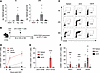 Figure 1
Early and sustained transgene presentation to T cells after recombinant AAV administration. (A) C57BL/6 mice (n = 5) were injected into the left tibialis anterior (TA) with 3 × 10¹⁰ vg of AAV1-CMV-luciferase. Left-side iliac (ili), inguinal (ing), and popliteal (pop) lymph nodes and right-side inguinal and popliteal lymph nodes (ndLN) were collected 1 or 24 hours after injection to perform AAV genome quantification by qPCR using transgene specific primers. (B) Experimental design for experiments in C–F: C57BL/6 mice were injected with 1 × 10¹0 vg of AAV into the left TA and subsequently received 1 × 10⁶ splenocytes from OT-I donor mice intravenously at the indicated time points. Spleen and left (draining; dLNs) and right (non-draining; ndLNs) pooled popliteal and inguinal lymph nodes were collected 18 hours after transfer and analyzed by flow cytometry. (C and D) As described in B, mice (n = 5) injected with AAV1-PGK-OVA257 received OT-I CD8+ T cells 4 hours, 24 hours, or 96 hours later. Representative dot plots (C) and kinetics (D) of the frequencies of CD69+CD25+ in live CD45.1+CD8+ OT-I T cells. (E and F) As described in B, mice (n = 4–10) were injected with either control AAV1-PGK-ESE or AAV1-PGK-cOVA (E) or different AAV serotypes (AAV1, AAV5, AAV8) encoding PGK-OVA257 (F) and received OT-I CD8+ T cells 24 hours later. Frequencies of CD69+CD25+ cells in live CD45.1+CD8+ OT-I T cells are represented. Each dot represents an individual mouse. Data are pooled from 2 (A), 3 (B–D), 1 (E), or 2 (F) independent experiments and are represented as mean ± SEM. Statistical significance was determined by 1-way ANOVA with Tukey’s multiple-comparison test: compared vs. ndLN (A and F) or dLN-AAV1-ESE (E), *P < 0.02 **P < 0.01, ****P < 0.0001; compared vs. AAV8, #P < 0.02, ####P < 0.0001 (F).
Unlike preformed protein antigens, transgenic proteins are produced only after AAV transduction, genome processing, and transgene expression. Consequently, presentation of transgene-derived peptides to CD8+ T cells is expected to occur later than that of exogenous proteins. To investigate this, we used the myotropic AAV1 vector, which exhibits limited liver transduction following intramuscular administration, thereby reducing the likelihood of liver-induced tolerance (30). The SIINFEKL peptide, placed under the control of the ubiquitous PGK promoter (AAV1-OVA257), was selected as the transgene, enabling CD8+ T cell activation to be analyzed independently of potential confounding factors such as anti-transgene CD4+ responses, antibody-mediated enhancement of CD8+ activation (23), or CD4+ regulatory T cells (31–33). After intramuscular administration of AAV1-OVA257, transgenic OVA257-specific CD8+ T cells (OT-I) were transferred intravenously at various time points. Their activation was assessed 18 hours later by the expression of the early activation markers CD25 and CD69 (Figure 1B). Strikingly, 24.5% ± 5.8% OT-I cells were activated in the dLNs as early as 24 hours after AAV administration, increasing to 35.3% ± 6.3% CD69+CD25+ at 96 hours (Figure 1, C and D). Activation was largely restricted to dLNs, with minimal activation detected in ndLNs and spleen.
We next asked whether this rapid presentation to T cells was specific to AAV1-OVA257 or represented a general feature of AAV vectors, regardless of encoded transgene or capsid serotype. First, we showed that the minimal OVA257 peptide transgene was not responsible for the rapid T cell activation. An AAV1 vector expressing cytoplasmic ovalbumin protein (cOVA), which is more representative of therapeutic intracellular transgenes and requires proteasomal processing before MHC class I presentation, also induced rapid OT-I activation in dLNs, whereas a control vector (AAV1-ESE) did not (Figure 1E). Furthermore, early CD8+ T cell activation was also observed in dLNs of mice injected with AAV5 and AAV8 vectors (Figure 1F). Notably, AAV8 induced greater activation in the dLNs and spleen compared with AAV1 and AAV5. As vector genome levels in the dLNs and spleen were comparable among serotypes (Supplemental Figure 1C; supplemental material available online with this article; https://doi.org/10.1172/jci.insight.202541DS1), these findings suggest serotype-dependent differences in APC transduction and/or transgene expression.
Finally, two additional control experiments were performed. First, we assessed the impact of injection volume: both lower (5 μL) and higher (100 μL) volume of AAV1-OVA257 resulted in similar OT-I activation in dLNs, suggesting that lymphatic drainage occurs independently of excessive muscle damage (Supplemental Figure 1A). Second, we verified that intact AAV particles were required for early presentation: before administration and OT-I transfer, AAV1-OVA257 vector and OVA257 peptide as control were heated to 95°C, a temperature that denatures AAV capsids and prevents transgene expression (34). As expected, heating the vector completely abolished OT-I activation, whereas heating the peptide had no effect (Supplemental Figure 1B).
Collectively, these findings demonstrate that AAV-encoded transgene is expressed and presented to T cells in the dLNs as early as 24 hours after administration.
Early transgene presentation in dLNs drives efficient T cell activation. Upregulation of early activation markers such as CD25 and CD69 reflects antigen encounter and does not necessarily indicate complete T cell activation and efficient clonal expansion. To assess this and follow initial T cell divisions, we tracked Violet Proliferation Dye–labeled (VPD-labeled) OT-I cells transferred 1 day before AAV1-OVA257 administration. As early as day 4, 71.4% ± 11.7% of OT-I cells had divided and accumulated in the dLNs and 44.5% ± 12.2% in the spleen, whereas few divided cells were present in the ndLNs and none in mice receiving the AAV1-ESE control vector (Figure 2, A and B). Importantly, in the absence of non-physiological OT-I cell transfer, we also detected at day 7 endogenous OVA257-specific CD8+ T cells that were effectively activated by AAV1-OVA257 and expanded (Figure 2, C and D). Under these conditions, 0.3% ± 0.1% of tetramer+ CD8+ T cells were found in the dLNs and 0.1% ± 0.05% in the spleen, versus 0% in ndLNs or after AAV1-ESE administration.
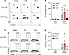 Figure 2
Early transgene presentation leads to efficient T cell activation. (A and B) C57BL/6 mice received intravenously 1 × 10⁶ VPD-labeled splenocytes from OT-I donor mice and 24 hours later were injected with 1 × 10¹⁰ vg of either AAV1-ESE (n = 6) or AAV1-OVA257 (n = 8) in the left TA (intramuscularly). On day 4, spleen, left dLNs (pooled popliteal and inguinal lymph nodes), and right ndLNs were collected and analyzed by flow cytometry. Representative dot plots (A) and frequencies (B) of VPDlo cells in live CD45.1+CD8+ OT-I T cells. (C and D) C57BL/6 mice were injected in the left TA with 1 × 10¹⁰ vg of either AAV1-ESE (n = 3) or AAV1-OVA257 (n = 5). On day 7, spleen, left dLNs, and right ndLNs were collected and analyzed by flow cytometry. Representative dot plots (C) and frequencies (D) of KbOVA257 tetramer+ CD44+ cells in live CD8+ cells. Each dot represents an individual mouse. Data are pooled from 3 (A and B) or 2 (C and D) independent experiments and are represented as mean ± SEM. Statistical significance was determined by 1-way ANOVA with Tukey’s multiple-comparison test: comparison vs. AAV1-ESE, **P < 0.01, ****P < 0.0001.
Next, we investigated whether AAV particles reaching the dLNs via lymphatic drainage — rather than active transport by DCs — were sufficient for efficient T cell activation. To this end, AAV1-OVA257 was injected intradermally into the top of the ear, a route previously shown to be at least as immunogenic as the intramuscular one (35). One hour later, the injection site was excised to ensure that only rapidly drained AAV particles remained (29). As expected, OT-I cells were activated in dLNs to a similar extent after ear excision versus in intact mice, confirming rapid lymphatic drainage of AAV particles (Figure 3A). No nonspecific activation was observed after ear excision upon administration of AAV1-ESE vector (Figure 3A). Critically, in a separate series of experiments without OT-I T cell transfer, we found that this early antigen encounter was sufficient to drive robust endogenous CD8+ T cell response (Figure 3B). As shown by tetramer staining, OVA257-specific CD8+ T cells were efficiently expanded despite removal of the injection site, with 5.2% ± 2.0% tetramer+ among CD8+ T cells, versus 7.0% ± 2.2% in intact mice, at day 14 after vector injection (Figure 3B). To test whether these CD8+ T cells induced after injection site removal were functional, an in vivo cytotoxic assay was performed by transfer of OVA257 peptide–loaded splenocytes in mice previously injected with AAV. Six weeks after AAV administration, VPD450hi OVA257 peptide–loaded splenocytes were equally rejected by mice with intact or excised injection sites (Figure 3, C and D).
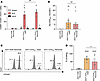 Figure 3
Early transgene presentation is sufficient to induce a specific CD8+ response. (A–D) C57BL/6 mice were injected intradermally in the top of the ear with 1 × 10¹⁰ vg of either AAV1-ESE or AAV1-OVA257. One hour later, the top of the ear was excised (Removed) or left intact (Intact). Different experiments were performed after this treatment and are represented in the corresponding panels. (A) Twenty-four hours after excision, mice (n = 3–5) received intravenously 1 × 10⁶ VPD-labeled splenocytes from OT-I donor mice and were euthanized 18 hours later. Spleen, superficial cervical lymph nodes (parotid and mandibular; dLNs), and popliteal and inguinal lymph nodes (ndLNs) were collected and analyzed by flow cytometry. Histogram showing the frequencies of CD69+CD25+ in live CD45.1+CD8+ OT-I T cells. (B) At 2 weeks after AAV, mice (n = 4–9) were bled to analyze KbOVA257 tetramer+ CD44+ cells in live CD8+ cells by flow cytometry. (C and D) Six weeks after ear excision, mice received through intravenous injection a mixture of unloaded and OVA257-loaded splenocytes (1 × 10⁷ total cells). Mice (n = 3–6) were euthanized 2 days later, and spleens were collected and analyzed by flow cytometry. (C) Representative histograms gated on live B220+ donor splenocytes. (D) Percentage of cytotoxicity. Each dot represents an individual mouse. Data are pooled from 3 (A–D) independent experiments and are represented as mean ± SEM. (A) Statistical significance was determined by 1-way ANOVA with Tukey’s multiple-comparison test: comparison vs. AAV1-ESE, *P < 0.1. (B–D) Statistical significance was determined by the Kruskal-Wallis test with Dunn’s multiple-comparison test, *P < 0.1, **P < 0.01, ****P < 0.0001.
Altogether, these results demonstrate that the AAV particles reaching the dLNs within the first hour after injection are sufficient to optimally prime and expand functional cytotoxic CD8+ T cells.
Muscle-specific expression reduces early antigen presentation and T cell activation. Given the importance of early antigen presentation for T cell priming, we wondered whether its reduction would be beneficial for limiting unwanted T cell responses. To this end, we sought to restrict transgene expression to muscle cells, thereby limiting expression in APCs and reducing early transgene-derived antigen presentation. We generated five AAV1 vectors encoding the OVA257 epitope, each driven by a different muscle-specific promoter. Their expression was confirmed in murine C2C12 myoblasts, by RT-qPCR analysis 24 hours after transduction (Figure 4A). Unexpectedly, transgene expression was also detected in DC2.4 murine dendritic cells (DCs) for all promoters. Expression levels were comparable for SPc5-12 and the ubiquitous PGK promoter, with SPc5-12 tending to induce a higher expression than the other muscle-specific promoters (Figure 4A). To determine whether this promoter leakiness in cultured DCs resulted in effective transgene presentation to CD8+ T cells, DC2.4 cells were transduced with the different AAV1 vectors at different MOI for 24 hours, then cocultured with OT-I T cells. Promoters enabled OT-I T cell activation with SPc5-12 and PGK promoters appearing stronger than human ACTA1 (hACTA1), desmin, or MHCK7, in agreement with RT-qPCR results (Figure 4B). These in vitro findings were confirmed in vivo using the previously described approach: intramuscular AAV administration followed by assessment of OT-I activation at 24 hours. Except desmin, for which the effect was not significant, all tested muscle-specific promoters supported antigen presentation in the dLNs, with SPc5-12 and PGK showing higher levels compared with hACTA1, desmin, and MHCK7 (Figure 4C). Comparable results were obtained in the spleen (Supplemental Figure 2A). This differential antigen presentation in lymphoid organs is not explained by differences in transduction efficiency or transgene expression, as equivalent levels of vector genome copy number and transgene mRNA were measured in muscle (Supplemental Figure 2, B and C). Overall, mRNA expression levels in DC2.4 cells were strongly correlated with antigen presentation both in vitro and in vivo (Supplemental Figure 2, D and E). Importantly, promoter leakiness resulting in elevated antigen presentation both in vitro and in vivo correlated with robust priming of transgene-specific CD8+ T cells. Thus, endogenous OVA257-specific CD8+ T cells were primed and expanded only following administration of SPc5-12 and PGK AAV1 vectors (Figure 4, D and E).
 Figure 4
Muscle-specific promoters reduce early antigen presentation and CD8+ T cell responses. (A) Murine myoblast C2C12 cells or dendritic DC2.4 cells were transduced by 1 × 10⁵ vg/cell of different AAV1 vectors encoding OVA257 under the control of indicated promoters (NT, not transduced). Transgene mRNA was quantified by specific RT-qPCR 24 hours after transduction. (B) DC2.4 cells were transduced with the indicated MOIs of the different AAV1-OVA257 vectors for 24 hours, and cocultured with CD8+ OT-I T cells for 18 hours. Expression of CD69/CD25 activation markers on CD8+ OT-I T cells was assessed by flow cytometry. (C) C57BL/6 mice (n = 8–10) were injected with 1 × 10¹⁰ vg of indicated AAV1-OVA257 vectors in the left TA 24 hours before receiving 1 × 10⁶ splenocytes from OT-I donor mice through intravenous injection. Left popliteal and inguinal dLNs were collected 18 hours later and analyzed by flow cytometry. Frequencies of CD69+CD25+ in live CD45.1+CD8+ OT-I T cells are represented. (D and E) C57BL/6 mice (n = 8) were injected in the left TA with indicated AAV1-OVA257 vectors and were bled at indicated time points to measure transgene-specific CD8+ T cells by flow cytometry. Representative dot plots (D) and kinetics (E) showing the frequencies of KbOVA257 tetramer+ CD44+ cells in live CD8+ cells. Each dot represents an independent experiment (A and B) or an individual mouse (C–E). Data are pooled from 2 (A and B) or 3 (C and E) independent experiments and are represented as mean ± SEM. Statistical significance was determined by the Kruskal-Wallis test with Dunn’s multiple-comparison test: comparison vs. Ctrl, *P < 0.1, ***P < 0.001, ****P < 0.0001; comparison vs. SPc5-12, #P < 0.01, ###P < 0.001.
Leakiness of SPc5-12 promoter was previously reported in plasmacytoid DCs (36). To limit its nonspecific activity in APCs, we inserted miR-142-3p target sequences into the SPc5-12 cassette, a strategy known to restrict transgene mRNA expression in hematopoietic cells (37). In vitro, this miR-based regulation reduced transgene mRNA expression in the DC2.4 cell line, as well as the activation of OT-I T cells (Figure 5, A and B). Consistently, in vivo transgene presentation at 24 hours was diminished, as evidenced by lower OT-I activation in mice receiving AAV1–SPc5-12–miR vector compared with those receiving AAV1–SPc5-12 vector (Figure 5C). This reduction could not be explained by decreased transgene expression in muscle, since equivalent levels were measured by RT-qPCR (Figure 5D). Importantly, in separate experiments, OVA257-specific CD8+ T cells were not detected in lymphoid organs (dLNs and spleen) 14 days after AAV1–SPc5-12–miR vector administration (Figure 5E), nor in blood during a 1-month follow-up (data not shown). Moreover, CD8+ T cell infiltration in the tibialis anterior was considerably reduced in mice injected with AAV1–SPc5-12–miR vector as compared with those injected with AAV1–SPc5-12 (Figure 5F). Consistently, muscle fiber integrity was preserved by the addition of the miR-142-3p target sequences. To further assess the fate of OVA257-specific CD8+ T cells, we transferred 1 × 10⁵ VPD-labeled OT-I cells prior to AAV vectors and followed their proliferation over time. In mice injected with miR-regulated AAV vectors, OT-I cells showed reduced proliferation (Figure 5, G and H), resulting from limited expansion in both dLNs and spleen at day 7 and no accumulation thereafter (Figure 5, I and J).
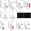 Figure 5
Enforced muscle-specific expression inhibits proper CD8+ T cell activation. (A) Transgene mRNA was quantified by specific RT-qPCR 24 hours after transduction of DC2.4 cells by 1 × 10⁵ vg/cell of indicated AAV1-OVA257 vectors. (B) DC2.4 cells were transduced with indicated MOIs of the different AAV1-OVA257 vectors for 24 hours, and cocultured with CD8+ OT-I T cells for 18 hours. Expression of CD69/CD25 on CD8+ OT-I T cells was assessed by flow cytometry. (C–J) C57BL/6 mice were injected intramuscularly in the left TA with 1 × 10¹⁰ vg of either AAV1–SPc5-12–OVA257, AAV1–SPc5-12–OVA257–miR, or an irrelevant vector (Irr) encoding murine SEAP under the control of the PGK promoter. (C and D) Twenty-four hours after AAV, mice (n = 5–10) received intravenously 1 × 10⁶ splenocytes from OT-I donor mice. dLNs and TA were collected 18 hours later. (C) Histogram showing the frequencies of CD69+CD25+ in live CD45.1+CD8+ OT-I T cells. (D) Transgene mRNA levels in the left TA were quantified by RT-qPCR. (E and F) Two weeks after AAV, dLNs and spleen were collected and analyzed by flow cytometry (n = 3). (E) Frequencies of KbOVA257 tetramer+ CD44+ cells in live CD8+ cells. (F) Immunofluorescence staining of left TA for CD8 (red), laminin (green), and DAPI (blue). Scale bars: 100 μM. (G–J) Mice (n = 3) received 1 × 10⁵ VPD-labeled splenocytes from OT-I donor mice before AAV injection in the left TA (1 × 10¹⁰ vg). Proliferation of OT-I cells in dLNs at day 7 (G and H) and frequency in spleen and dLNs at indicated time points (I and J) were assessed by flow cytometry. Each dot represents an individual mouse. Data are pooled from 3 (A, C, and D) or 1 (B, E–J) independent experiments and are represented as mean ± SEM. Statistical analysis: (A and E) Kruskal-Wallis test with Dunn’s multiple-comparison test: vs. Ctrl or Irr, *P < 0.1, **P < 0.01. (C, D, and H) Mann-Whitney U test: *P < 0.1, ***P > 0,0001. (I and J) 2-way ANOVA: vs. SPc5-12; *P < 0.1, **P < 0.01.
Altogether, our results indicate that enforcing muscle-specific expression reduces early transgene presentation and prevents efficient CD8+ T cell activation.


