
Clinical Research and Public HealthInfectious diseasePulmonology
Open Access |  10.1172/jci.insight.199983
Pulmonary fibrosis after COVID-19 is characterized by airway abnormalities and elevated club cell secretory protein-16
Matthew R. Baldwin,1,2 Ansley E. Jones,1 David Zhang,1 Chandan Gurung,1 Zain Khan,3 Anjali Saqi,4 Xuehan Yang,3 Ying Wei,3 Renu Nandakumar,2 Scarlett O. Murphy,1 Claire F. McGroder,1 Faisal Shaikh,1 Selim Arcasoy,1 Luke Benvenuto,1 Harpreet Grewal,1 Benjamin M. Smith,1,5 Eric A. Hoffman,6 Agnes C.Y. Yuen,7 Parteek Johal,7 Christopher Carlsten,7 Christopher J. Ryerson,7 J. Brent Richards,8,9 Alyson W. Wong,7 Tomoko Nakanishi,10,11,12,13 Aditi S. Shah,7 and Christine Kim Garcia1,15
1Department of Medicine, Division of Pulmonary, Allergy, and Critical Care, Columbia University Vagelos College of Physicians and Surgeons, New York, New York, USA.
2Irving Institute for Clinical and Translational Research, Columbia University Medical Center, New York, New York, USA.
3Department of Biostatistics, Columbia University Mailman School of Public Health, New York, New York, USA.
4Department of Pathology, Columbia University Vagelos College of Physicians and Surgeons, New York, New York, USA.
5Department of Medicine, McGill University, Montreal, Quebec, Canada.
6Department of Radiology, Internal Medicine, and Biomedical Engineering, University of Iowa, Iowa City, Iowa.
7Division of Respiratory Medicine, Department of Medicine, University of British Columbia, Vancouver, British Columbia, Canada.
8Departments of Medicine, Human Genetics, Epidemiology and Biostatistics, McGill University, Montréal, Quebec, Canada.
9Department of Twin Research, King’s College London, London, United Kingdom.
10Department of Human Genetics, and
11Lady Davis Institute, Jewish General Hospital, McGill University, Montréal, Quebec, Canada.
12Kyoto-McGill International Collaborative Program in Genomic Medicine, Graduate School of Medicine, Kyoto University, Kyoto, Japan.
13Department of Genome Informatics, Graduate School of Medicine, the University of Tokyo, Tokyo, Japan.
14Research Fellow, Japan Society for the Promotion of Science, Tokyo, Japan.
15Columbia Precision Medicine Initiative, Columbia University Vagelos College of Physicians and Surgeons, New York, New York, USA.
Address correspondence to: Matthew R. Baldwin, 622 West 168th Street, PH8E, Room 101, New York, New York, 10032, USA. Email: mrb45@cumc.columbia.edu.
Authorship note: AEJ, DZ, and CG have been designated as co–second authors.
Find articles by
Baldwin, M.
in:
PubMed
|
Google Scholar
|

1Department of Medicine, Division of Pulmonary, Allergy, and Critical Care, Columbia University Vagelos College of Physicians and Surgeons, New York, New York, USA.
2Irving Institute for Clinical and Translational Research, Columbia University Medical Center, New York, New York, USA.
3Department of Biostatistics, Columbia University Mailman School of Public Health, New York, New York, USA.
4Department of Pathology, Columbia University Vagelos College of Physicians and Surgeons, New York, New York, USA.
5Department of Medicine, McGill University, Montreal, Quebec, Canada.
6Department of Radiology, Internal Medicine, and Biomedical Engineering, University of Iowa, Iowa City, Iowa.
7Division of Respiratory Medicine, Department of Medicine, University of British Columbia, Vancouver, British Columbia, Canada.
8Departments of Medicine, Human Genetics, Epidemiology and Biostatistics, McGill University, Montréal, Quebec, Canada.
9Department of Twin Research, King’s College London, London, United Kingdom.
10Department of Human Genetics, and
11Lady Davis Institute, Jewish General Hospital, McGill University, Montréal, Quebec, Canada.
12Kyoto-McGill International Collaborative Program in Genomic Medicine, Graduate School of Medicine, Kyoto University, Kyoto, Japan.
13Department of Genome Informatics, Graduate School of Medicine, the University of Tokyo, Tokyo, Japan.
14Research Fellow, Japan Society for the Promotion of Science, Tokyo, Japan.
15Columbia Precision Medicine Initiative, Columbia University Vagelos College of Physicians and Surgeons, New York, New York, USA.
Address correspondence to: Matthew R. Baldwin, 622 West 168th Street, PH8E, Room 101, New York, New York, 10032, USA. Email: mrb45@cumc.columbia.edu.
Authorship note: AEJ, DZ, and CG have been designated as co–second authors.
Find articles by Jones, A. in: PubMed | Google Scholar
1Department of Medicine, Division of Pulmonary, Allergy, and Critical Care, Columbia University Vagelos College of Physicians and Surgeons, New York, New York, USA.
2Irving Institute for Clinical and Translational Research, Columbia University Medical Center, New York, New York, USA.
3Department of Biostatistics, Columbia University Mailman School of Public Health, New York, New York, USA.
4Department of Pathology, Columbia University Vagelos College of Physicians and Surgeons, New York, New York, USA.
5Department of Medicine, McGill University, Montreal, Quebec, Canada.
6Department of Radiology, Internal Medicine, and Biomedical Engineering, University of Iowa, Iowa City, Iowa.
7Division of Respiratory Medicine, Department of Medicine, University of British Columbia, Vancouver, British Columbia, Canada.
8Departments of Medicine, Human Genetics, Epidemiology and Biostatistics, McGill University, Montréal, Quebec, Canada.
9Department of Twin Research, King’s College London, London, United Kingdom.
10Department of Human Genetics, and
11Lady Davis Institute, Jewish General Hospital, McGill University, Montréal, Quebec, Canada.
12Kyoto-McGill International Collaborative Program in Genomic Medicine, Graduate School of Medicine, Kyoto University, Kyoto, Japan.
13Department of Genome Informatics, Graduate School of Medicine, the University of Tokyo, Tokyo, Japan.
14Research Fellow, Japan Society for the Promotion of Science, Tokyo, Japan.
15Columbia Precision Medicine Initiative, Columbia University Vagelos College of Physicians and Surgeons, New York, New York, USA.
Address correspondence to: Matthew R. Baldwin, 622 West 168th Street, PH8E, Room 101, New York, New York, 10032, USA. Email: mrb45@cumc.columbia.edu.
Authorship note: AEJ, DZ, and CG have been designated as co–second authors.
Find articles by
Zhang, D.
in:
PubMed
|
Google Scholar
|
1Department of Medicine, Division of Pulmonary, Allergy, and Critical Care, Columbia University Vagelos College of Physicians and Surgeons, New York, New York, USA.
2Irving Institute for Clinical and Translational Research, Columbia University Medical Center, New York, New York, USA.
3Department of Biostatistics, Columbia University Mailman School of Public Health, New York, New York, USA.
4Department of Pathology, Columbia University Vagelos College of Physicians and Surgeons, New York, New York, USA.
5Department of Medicine, McGill University, Montreal, Quebec, Canada.
6Department of Radiology, Internal Medicine, and Biomedical Engineering, University of Iowa, Iowa City, Iowa.
7Division of Respiratory Medicine, Department of Medicine, University of British Columbia, Vancouver, British Columbia, Canada.
8Departments of Medicine, Human Genetics, Epidemiology and Biostatistics, McGill University, Montréal, Quebec, Canada.
9Department of Twin Research, King’s College London, London, United Kingdom.
10Department of Human Genetics, and
11Lady Davis Institute, Jewish General Hospital, McGill University, Montréal, Quebec, Canada.
12Kyoto-McGill International Collaborative Program in Genomic Medicine, Graduate School of Medicine, Kyoto University, Kyoto, Japan.
13Department of Genome Informatics, Graduate School of Medicine, the University of Tokyo, Tokyo, Japan.
14Research Fellow, Japan Society for the Promotion of Science, Tokyo, Japan.
15Columbia Precision Medicine Initiative, Columbia University Vagelos College of Physicians and Surgeons, New York, New York, USA.
Address correspondence to: Matthew R. Baldwin, 622 West 168th Street, PH8E, Room 101, New York, New York, 10032, USA. Email: mrb45@cumc.columbia.edu.
Authorship note: AEJ, DZ, and CG have been designated as co–second authors.
Find articles by Gurung, C. in: PubMed | Google Scholar
1Department of Medicine, Division of Pulmonary, Allergy, and Critical Care, Columbia University Vagelos College of Physicians and Surgeons, New York, New York, USA.
2Irving Institute for Clinical and Translational Research, Columbia University Medical Center, New York, New York, USA.
3Department of Biostatistics, Columbia University Mailman School of Public Health, New York, New York, USA.
4Department of Pathology, Columbia University Vagelos College of Physicians and Surgeons, New York, New York, USA.
5Department of Medicine, McGill University, Montreal, Quebec, Canada.
6Department of Radiology, Internal Medicine, and Biomedical Engineering, University of Iowa, Iowa City, Iowa.
7Division of Respiratory Medicine, Department of Medicine, University of British Columbia, Vancouver, British Columbia, Canada.
8Departments of Medicine, Human Genetics, Epidemiology and Biostatistics, McGill University, Montréal, Quebec, Canada.
9Department of Twin Research, King’s College London, London, United Kingdom.
10Department of Human Genetics, and
11Lady Davis Institute, Jewish General Hospital, McGill University, Montréal, Quebec, Canada.
12Kyoto-McGill International Collaborative Program in Genomic Medicine, Graduate School of Medicine, Kyoto University, Kyoto, Japan.
13Department of Genome Informatics, Graduate School of Medicine, the University of Tokyo, Tokyo, Japan.
14Research Fellow, Japan Society for the Promotion of Science, Tokyo, Japan.
15Columbia Precision Medicine Initiative, Columbia University Vagelos College of Physicians and Surgeons, New York, New York, USA.
Address correspondence to: Matthew R. Baldwin, 622 West 168th Street, PH8E, Room 101, New York, New York, 10032, USA. Email: mrb45@cumc.columbia.edu.
Authorship note: AEJ, DZ, and CG have been designated as co–second authors.
Find articles by Khan, Z. in: PubMed | Google Scholar
1Department of Medicine, Division of Pulmonary, Allergy, and Critical Care, Columbia University Vagelos College of Physicians and Surgeons, New York, New York, USA.
2Irving Institute for Clinical and Translational Research, Columbia University Medical Center, New York, New York, USA.
3Department of Biostatistics, Columbia University Mailman School of Public Health, New York, New York, USA.
4Department of Pathology, Columbia University Vagelos College of Physicians and Surgeons, New York, New York, USA.
5Department of Medicine, McGill University, Montreal, Quebec, Canada.
6Department of Radiology, Internal Medicine, and Biomedical Engineering, University of Iowa, Iowa City, Iowa.
7Division of Respiratory Medicine, Department of Medicine, University of British Columbia, Vancouver, British Columbia, Canada.
8Departments of Medicine, Human Genetics, Epidemiology and Biostatistics, McGill University, Montréal, Quebec, Canada.
9Department of Twin Research, King’s College London, London, United Kingdom.
10Department of Human Genetics, and
11Lady Davis Institute, Jewish General Hospital, McGill University, Montréal, Quebec, Canada.
12Kyoto-McGill International Collaborative Program in Genomic Medicine, Graduate School of Medicine, Kyoto University, Kyoto, Japan.
13Department of Genome Informatics, Graduate School of Medicine, the University of Tokyo, Tokyo, Japan.
14Research Fellow, Japan Society for the Promotion of Science, Tokyo, Japan.
15Columbia Precision Medicine Initiative, Columbia University Vagelos College of Physicians and Surgeons, New York, New York, USA.
Address correspondence to: Matthew R. Baldwin, 622 West 168th Street, PH8E, Room 101, New York, New York, 10032, USA. Email: mrb45@cumc.columbia.edu.
Authorship note: AEJ, DZ, and CG have been designated as co–second authors.
Find articles by Saqi, A. in: PubMed | Google Scholar
1Department of Medicine, Division of Pulmonary, Allergy, and Critical Care, Columbia University Vagelos College of Physicians and Surgeons, New York, New York, USA.
2Irving Institute for Clinical and Translational Research, Columbia University Medical Center, New York, New York, USA.
3Department of Biostatistics, Columbia University Mailman School of Public Health, New York, New York, USA.
4Department of Pathology, Columbia University Vagelos College of Physicians and Surgeons, New York, New York, USA.
5Department of Medicine, McGill University, Montreal, Quebec, Canada.
6Department of Radiology, Internal Medicine, and Biomedical Engineering, University of Iowa, Iowa City, Iowa.
7Division of Respiratory Medicine, Department of Medicine, University of British Columbia, Vancouver, British Columbia, Canada.
8Departments of Medicine, Human Genetics, Epidemiology and Biostatistics, McGill University, Montréal, Quebec, Canada.
9Department of Twin Research, King’s College London, London, United Kingdom.
10Department of Human Genetics, and
11Lady Davis Institute, Jewish General Hospital, McGill University, Montréal, Quebec, Canada.
12Kyoto-McGill International Collaborative Program in Genomic Medicine, Graduate School of Medicine, Kyoto University, Kyoto, Japan.
13Department of Genome Informatics, Graduate School of Medicine, the University of Tokyo, Tokyo, Japan.
14Research Fellow, Japan Society for the Promotion of Science, Tokyo, Japan.
15Columbia Precision Medicine Initiative, Columbia University Vagelos College of Physicians and Surgeons, New York, New York, USA.
Address correspondence to: Matthew R. Baldwin, 622 West 168th Street, PH8E, Room 101, New York, New York, 10032, USA. Email: mrb45@cumc.columbia.edu.
Authorship note: AEJ, DZ, and CG have been designated as co–second authors.
Find articles by Yang, X. in: PubMed | Google Scholar
1Department of Medicine, Division of Pulmonary, Allergy, and Critical Care, Columbia University Vagelos College of Physicians and Surgeons, New York, New York, USA.
2Irving Institute for Clinical and Translational Research, Columbia University Medical Center, New York, New York, USA.
3Department of Biostatistics, Columbia University Mailman School of Public Health, New York, New York, USA.
4Department of Pathology, Columbia University Vagelos College of Physicians and Surgeons, New York, New York, USA.
5Department of Medicine, McGill University, Montreal, Quebec, Canada.
6Department of Radiology, Internal Medicine, and Biomedical Engineering, University of Iowa, Iowa City, Iowa.
7Division of Respiratory Medicine, Department of Medicine, University of British Columbia, Vancouver, British Columbia, Canada.
8Departments of Medicine, Human Genetics, Epidemiology and Biostatistics, McGill University, Montréal, Quebec, Canada.
9Department of Twin Research, King’s College London, London, United Kingdom.
10Department of Human Genetics, and
11Lady Davis Institute, Jewish General Hospital, McGill University, Montréal, Quebec, Canada.
12Kyoto-McGill International Collaborative Program in Genomic Medicine, Graduate School of Medicine, Kyoto University, Kyoto, Japan.
13Department of Genome Informatics, Graduate School of Medicine, the University of Tokyo, Tokyo, Japan.
14Research Fellow, Japan Society for the Promotion of Science, Tokyo, Japan.
15Columbia Precision Medicine Initiative, Columbia University Vagelos College of Physicians and Surgeons, New York, New York, USA.
Address correspondence to: Matthew R. Baldwin, 622 West 168th Street, PH8E, Room 101, New York, New York, 10032, USA. Email: mrb45@cumc.columbia.edu.
Authorship note: AEJ, DZ, and CG have been designated as co–second authors.
Find articles by Wei, Y. in: PubMed | Google Scholar
1Department of Medicine, Division of Pulmonary, Allergy, and Critical Care, Columbia University Vagelos College of Physicians and Surgeons, New York, New York, USA.
2Irving Institute for Clinical and Translational Research, Columbia University Medical Center, New York, New York, USA.
3Department of Biostatistics, Columbia University Mailman School of Public Health, New York, New York, USA.
4Department of Pathology, Columbia University Vagelos College of Physicians and Surgeons, New York, New York, USA.
5Department of Medicine, McGill University, Montreal, Quebec, Canada.
6Department of Radiology, Internal Medicine, and Biomedical Engineering, University of Iowa, Iowa City, Iowa.
7Division of Respiratory Medicine, Department of Medicine, University of British Columbia, Vancouver, British Columbia, Canada.
8Departments of Medicine, Human Genetics, Epidemiology and Biostatistics, McGill University, Montréal, Quebec, Canada.
9Department of Twin Research, King’s College London, London, United Kingdom.
10Department of Human Genetics, and
11Lady Davis Institute, Jewish General Hospital, McGill University, Montréal, Quebec, Canada.
12Kyoto-McGill International Collaborative Program in Genomic Medicine, Graduate School of Medicine, Kyoto University, Kyoto, Japan.
13Department of Genome Informatics, Graduate School of Medicine, the University of Tokyo, Tokyo, Japan.
14Research Fellow, Japan Society for the Promotion of Science, Tokyo, Japan.
15Columbia Precision Medicine Initiative, Columbia University Vagelos College of Physicians and Surgeons, New York, New York, USA.
Address correspondence to: Matthew R. Baldwin, 622 West 168th Street, PH8E, Room 101, New York, New York, 10032, USA. Email: mrb45@cumc.columbia.edu.
Authorship note: AEJ, DZ, and CG have been designated as co–second authors.
Find articles by Nandakumar, R. in: PubMed | Google Scholar
1Department of Medicine, Division of Pulmonary, Allergy, and Critical Care, Columbia University Vagelos College of Physicians and Surgeons, New York, New York, USA.
2Irving Institute for Clinical and Translational Research, Columbia University Medical Center, New York, New York, USA.
3Department of Biostatistics, Columbia University Mailman School of Public Health, New York, New York, USA.
4Department of Pathology, Columbia University Vagelos College of Physicians and Surgeons, New York, New York, USA.
5Department of Medicine, McGill University, Montreal, Quebec, Canada.
6Department of Radiology, Internal Medicine, and Biomedical Engineering, University of Iowa, Iowa City, Iowa.
7Division of Respiratory Medicine, Department of Medicine, University of British Columbia, Vancouver, British Columbia, Canada.
8Departments of Medicine, Human Genetics, Epidemiology and Biostatistics, McGill University, Montréal, Quebec, Canada.
9Department of Twin Research, King’s College London, London, United Kingdom.
10Department of Human Genetics, and
11Lady Davis Institute, Jewish General Hospital, McGill University, Montréal, Quebec, Canada.
12Kyoto-McGill International Collaborative Program in Genomic Medicine, Graduate School of Medicine, Kyoto University, Kyoto, Japan.
13Department of Genome Informatics, Graduate School of Medicine, the University of Tokyo, Tokyo, Japan.
14Research Fellow, Japan Society for the Promotion of Science, Tokyo, Japan.
15Columbia Precision Medicine Initiative, Columbia University Vagelos College of Physicians and Surgeons, New York, New York, USA.
Address correspondence to: Matthew R. Baldwin, 622 West 168th Street, PH8E, Room 101, New York, New York, 10032, USA. Email: mrb45@cumc.columbia.edu.
Authorship note: AEJ, DZ, and CG have been designated as co–second authors.
Find articles by Murphy, S. in: PubMed | Google Scholar
1Department of Medicine, Division of Pulmonary, Allergy, and Critical Care, Columbia University Vagelos College of Physicians and Surgeons, New York, New York, USA.
2Irving Institute for Clinical and Translational Research, Columbia University Medical Center, New York, New York, USA.
3Department of Biostatistics, Columbia University Mailman School of Public Health, New York, New York, USA.
4Department of Pathology, Columbia University Vagelos College of Physicians and Surgeons, New York, New York, USA.
5Department of Medicine, McGill University, Montreal, Quebec, Canada.
6Department of Radiology, Internal Medicine, and Biomedical Engineering, University of Iowa, Iowa City, Iowa.
7Division of Respiratory Medicine, Department of Medicine, University of British Columbia, Vancouver, British Columbia, Canada.
8Departments of Medicine, Human Genetics, Epidemiology and Biostatistics, McGill University, Montréal, Quebec, Canada.
9Department of Twin Research, King’s College London, London, United Kingdom.
10Department of Human Genetics, and
11Lady Davis Institute, Jewish General Hospital, McGill University, Montréal, Quebec, Canada.
12Kyoto-McGill International Collaborative Program in Genomic Medicine, Graduate School of Medicine, Kyoto University, Kyoto, Japan.
13Department of Genome Informatics, Graduate School of Medicine, the University of Tokyo, Tokyo, Japan.
14Research Fellow, Japan Society for the Promotion of Science, Tokyo, Japan.
15Columbia Precision Medicine Initiative, Columbia University Vagelos College of Physicians and Surgeons, New York, New York, USA.
Address correspondence to: Matthew R. Baldwin, 622 West 168th Street, PH8E, Room 101, New York, New York, 10032, USA. Email: mrb45@cumc.columbia.edu.
Authorship note: AEJ, DZ, and CG have been designated as co–second authors.
Find articles by McGroder, C. in: PubMed | Google Scholar
1Department of Medicine, Division of Pulmonary, Allergy, and Critical Care, Columbia University Vagelos College of Physicians and Surgeons, New York, New York, USA.
2Irving Institute for Clinical and Translational Research, Columbia University Medical Center, New York, New York, USA.
3Department of Biostatistics, Columbia University Mailman School of Public Health, New York, New York, USA.
4Department of Pathology, Columbia University Vagelos College of Physicians and Surgeons, New York, New York, USA.
5Department of Medicine, McGill University, Montreal, Quebec, Canada.
6Department of Radiology, Internal Medicine, and Biomedical Engineering, University of Iowa, Iowa City, Iowa.
7Division of Respiratory Medicine, Department of Medicine, University of British Columbia, Vancouver, British Columbia, Canada.
8Departments of Medicine, Human Genetics, Epidemiology and Biostatistics, McGill University, Montréal, Quebec, Canada.
9Department of Twin Research, King’s College London, London, United Kingdom.
10Department of Human Genetics, and
11Lady Davis Institute, Jewish General Hospital, McGill University, Montréal, Quebec, Canada.
12Kyoto-McGill International Collaborative Program in Genomic Medicine, Graduate School of Medicine, Kyoto University, Kyoto, Japan.
13Department of Genome Informatics, Graduate School of Medicine, the University of Tokyo, Tokyo, Japan.
14Research Fellow, Japan Society for the Promotion of Science, Tokyo, Japan.
15Columbia Precision Medicine Initiative, Columbia University Vagelos College of Physicians and Surgeons, New York, New York, USA.
Address correspondence to: Matthew R. Baldwin, 622 West 168th Street, PH8E, Room 101, New York, New York, 10032, USA. Email: mrb45@cumc.columbia.edu.
Authorship note: AEJ, DZ, and CG have been designated as co–second authors.
Find articles by Shaikh, F. in: PubMed | Google Scholar
1Department of Medicine, Division of Pulmonary, Allergy, and Critical Care, Columbia University Vagelos College of Physicians and Surgeons, New York, New York, USA.
2Irving Institute for Clinical and Translational Research, Columbia University Medical Center, New York, New York, USA.
3Department of Biostatistics, Columbia University Mailman School of Public Health, New York, New York, USA.
4Department of Pathology, Columbia University Vagelos College of Physicians and Surgeons, New York, New York, USA.
5Department of Medicine, McGill University, Montreal, Quebec, Canada.
6Department of Radiology, Internal Medicine, and Biomedical Engineering, University of Iowa, Iowa City, Iowa.
7Division of Respiratory Medicine, Department of Medicine, University of British Columbia, Vancouver, British Columbia, Canada.
8Departments of Medicine, Human Genetics, Epidemiology and Biostatistics, McGill University, Montréal, Quebec, Canada.
9Department of Twin Research, King’s College London, London, United Kingdom.
10Department of Human Genetics, and
11Lady Davis Institute, Jewish General Hospital, McGill University, Montréal, Quebec, Canada.
12Kyoto-McGill International Collaborative Program in Genomic Medicine, Graduate School of Medicine, Kyoto University, Kyoto, Japan.
13Department of Genome Informatics, Graduate School of Medicine, the University of Tokyo, Tokyo, Japan.
14Research Fellow, Japan Society for the Promotion of Science, Tokyo, Japan.
15Columbia Precision Medicine Initiative, Columbia University Vagelos College of Physicians and Surgeons, New York, New York, USA.
Address correspondence to: Matthew R. Baldwin, 622 West 168th Street, PH8E, Room 101, New York, New York, 10032, USA. Email: mrb45@cumc.columbia.edu.
Authorship note: AEJ, DZ, and CG have been designated as co–second authors.
Find articles by
Arcasoy, S.
in:
PubMed
|
Google Scholar
|
1Department of Medicine, Division of Pulmonary, Allergy, and Critical Care, Columbia University Vagelos College of Physicians and Surgeons, New York, New York, USA.
2Irving Institute for Clinical and Translational Research, Columbia University Medical Center, New York, New York, USA.
3Department of Biostatistics, Columbia University Mailman School of Public Health, New York, New York, USA.
4Department of Pathology, Columbia University Vagelos College of Physicians and Surgeons, New York, New York, USA.
5Department of Medicine, McGill University, Montreal, Quebec, Canada.
6Department of Radiology, Internal Medicine, and Biomedical Engineering, University of Iowa, Iowa City, Iowa.
7Division of Respiratory Medicine, Department of Medicine, University of British Columbia, Vancouver, British Columbia, Canada.
8Departments of Medicine, Human Genetics, Epidemiology and Biostatistics, McGill University, Montréal, Quebec, Canada.
9Department of Twin Research, King’s College London, London, United Kingdom.
10Department of Human Genetics, and
11Lady Davis Institute, Jewish General Hospital, McGill University, Montréal, Quebec, Canada.
12Kyoto-McGill International Collaborative Program in Genomic Medicine, Graduate School of Medicine, Kyoto University, Kyoto, Japan.
13Department of Genome Informatics, Graduate School of Medicine, the University of Tokyo, Tokyo, Japan.
14Research Fellow, Japan Society for the Promotion of Science, Tokyo, Japan.
15Columbia Precision Medicine Initiative, Columbia University Vagelos College of Physicians and Surgeons, New York, New York, USA.
Address correspondence to: Matthew R. Baldwin, 622 West 168th Street, PH8E, Room 101, New York, New York, 10032, USA. Email: mrb45@cumc.columbia.edu.
Authorship note: AEJ, DZ, and CG have been designated as co–second authors.
Find articles by
Benvenuto, L.
in:
PubMed
|
Google Scholar
|
1Department of Medicine, Division of Pulmonary, Allergy, and Critical Care, Columbia University Vagelos College of Physicians and Surgeons, New York, New York, USA.
2Irving Institute for Clinical and Translational Research, Columbia University Medical Center, New York, New York, USA.
3Department of Biostatistics, Columbia University Mailman School of Public Health, New York, New York, USA.
4Department of Pathology, Columbia University Vagelos College of Physicians and Surgeons, New York, New York, USA.
5Department of Medicine, McGill University, Montreal, Quebec, Canada.
6Department of Radiology, Internal Medicine, and Biomedical Engineering, University of Iowa, Iowa City, Iowa.
7Division of Respiratory Medicine, Department of Medicine, University of British Columbia, Vancouver, British Columbia, Canada.
8Departments of Medicine, Human Genetics, Epidemiology and Biostatistics, McGill University, Montréal, Quebec, Canada.
9Department of Twin Research, King’s College London, London, United Kingdom.
10Department of Human Genetics, and
11Lady Davis Institute, Jewish General Hospital, McGill University, Montréal, Quebec, Canada.
12Kyoto-McGill International Collaborative Program in Genomic Medicine, Graduate School of Medicine, Kyoto University, Kyoto, Japan.
13Department of Genome Informatics, Graduate School of Medicine, the University of Tokyo, Tokyo, Japan.
14Research Fellow, Japan Society for the Promotion of Science, Tokyo, Japan.
15Columbia Precision Medicine Initiative, Columbia University Vagelos College of Physicians and Surgeons, New York, New York, USA.
Address correspondence to: Matthew R. Baldwin, 622 West 168th Street, PH8E, Room 101, New York, New York, 10032, USA. Email: mrb45@cumc.columbia.edu.
Authorship note: AEJ, DZ, and CG have been designated as co–second authors.
Find articles by Grewal, H. in: PubMed | Google Scholar
1Department of Medicine, Division of Pulmonary, Allergy, and Critical Care, Columbia University Vagelos College of Physicians and Surgeons, New York, New York, USA.
2Irving Institute for Clinical and Translational Research, Columbia University Medical Center, New York, New York, USA.
3Department of Biostatistics, Columbia University Mailman School of Public Health, New York, New York, USA.
4Department of Pathology, Columbia University Vagelos College of Physicians and Surgeons, New York, New York, USA.
5Department of Medicine, McGill University, Montreal, Quebec, Canada.
6Department of Radiology, Internal Medicine, and Biomedical Engineering, University of Iowa, Iowa City, Iowa.
7Division of Respiratory Medicine, Department of Medicine, University of British Columbia, Vancouver, British Columbia, Canada.
8Departments of Medicine, Human Genetics, Epidemiology and Biostatistics, McGill University, Montréal, Quebec, Canada.
9Department of Twin Research, King’s College London, London, United Kingdom.
10Department of Human Genetics, and
11Lady Davis Institute, Jewish General Hospital, McGill University, Montréal, Quebec, Canada.
12Kyoto-McGill International Collaborative Program in Genomic Medicine, Graduate School of Medicine, Kyoto University, Kyoto, Japan.
13Department of Genome Informatics, Graduate School of Medicine, the University of Tokyo, Tokyo, Japan.
14Research Fellow, Japan Society for the Promotion of Science, Tokyo, Japan.
15Columbia Precision Medicine Initiative, Columbia University Vagelos College of Physicians and Surgeons, New York, New York, USA.
Address correspondence to: Matthew R. Baldwin, 622 West 168th Street, PH8E, Room 101, New York, New York, 10032, USA. Email: mrb45@cumc.columbia.edu.
Authorship note: AEJ, DZ, and CG have been designated as co–second authors.
Find articles by Smith, B. in: PubMed | Google Scholar
1Department of Medicine, Division of Pulmonary, Allergy, and Critical Care, Columbia University Vagelos College of Physicians and Surgeons, New York, New York, USA.
2Irving Institute for Clinical and Translational Research, Columbia University Medical Center, New York, New York, USA.
3Department of Biostatistics, Columbia University Mailman School of Public Health, New York, New York, USA.
4Department of Pathology, Columbia University Vagelos College of Physicians and Surgeons, New York, New York, USA.
5Department of Medicine, McGill University, Montreal, Quebec, Canada.
6Department of Radiology, Internal Medicine, and Biomedical Engineering, University of Iowa, Iowa City, Iowa.
7Division of Respiratory Medicine, Department of Medicine, University of British Columbia, Vancouver, British Columbia, Canada.
8Departments of Medicine, Human Genetics, Epidemiology and Biostatistics, McGill University, Montréal, Quebec, Canada.
9Department of Twin Research, King’s College London, London, United Kingdom.
10Department of Human Genetics, and
11Lady Davis Institute, Jewish General Hospital, McGill University, Montréal, Quebec, Canada.
12Kyoto-McGill International Collaborative Program in Genomic Medicine, Graduate School of Medicine, Kyoto University, Kyoto, Japan.
13Department of Genome Informatics, Graduate School of Medicine, the University of Tokyo, Tokyo, Japan.
14Research Fellow, Japan Society for the Promotion of Science, Tokyo, Japan.
15Columbia Precision Medicine Initiative, Columbia University Vagelos College of Physicians and Surgeons, New York, New York, USA.
Address correspondence to: Matthew R. Baldwin, 622 West 168th Street, PH8E, Room 101, New York, New York, 10032, USA. Email: mrb45@cumc.columbia.edu.
Authorship note: AEJ, DZ, and CG have been designated as co–second authors.
Find articles by
Hoffman, E.
in:
PubMed
|
Google Scholar
|
1Department of Medicine, Division of Pulmonary, Allergy, and Critical Care, Columbia University Vagelos College of Physicians and Surgeons, New York, New York, USA.
2Irving Institute for Clinical and Translational Research, Columbia University Medical Center, New York, New York, USA.
3Department of Biostatistics, Columbia University Mailman School of Public Health, New York, New York, USA.
4Department of Pathology, Columbia University Vagelos College of Physicians and Surgeons, New York, New York, USA.
5Department of Medicine, McGill University, Montreal, Quebec, Canada.
6Department of Radiology, Internal Medicine, and Biomedical Engineering, University of Iowa, Iowa City, Iowa.
7Division of Respiratory Medicine, Department of Medicine, University of British Columbia, Vancouver, British Columbia, Canada.
8Departments of Medicine, Human Genetics, Epidemiology and Biostatistics, McGill University, Montréal, Quebec, Canada.
9Department of Twin Research, King’s College London, London, United Kingdom.
10Department of Human Genetics, and
11Lady Davis Institute, Jewish General Hospital, McGill University, Montréal, Quebec, Canada.
12Kyoto-McGill International Collaborative Program in Genomic Medicine, Graduate School of Medicine, Kyoto University, Kyoto, Japan.
13Department of Genome Informatics, Graduate School of Medicine, the University of Tokyo, Tokyo, Japan.
14Research Fellow, Japan Society for the Promotion of Science, Tokyo, Japan.
15Columbia Precision Medicine Initiative, Columbia University Vagelos College of Physicians and Surgeons, New York, New York, USA.
Address correspondence to: Matthew R. Baldwin, 622 West 168th Street, PH8E, Room 101, New York, New York, 10032, USA. Email: mrb45@cumc.columbia.edu.
Authorship note: AEJ, DZ, and CG have been designated as co–second authors.
Find articles by Yuen, A. in: PubMed | Google Scholar
1Department of Medicine, Division of Pulmonary, Allergy, and Critical Care, Columbia University Vagelos College of Physicians and Surgeons, New York, New York, USA.
2Irving Institute for Clinical and Translational Research, Columbia University Medical Center, New York, New York, USA.
3Department of Biostatistics, Columbia University Mailman School of Public Health, New York, New York, USA.
4Department of Pathology, Columbia University Vagelos College of Physicians and Surgeons, New York, New York, USA.
5Department of Medicine, McGill University, Montreal, Quebec, Canada.
6Department of Radiology, Internal Medicine, and Biomedical Engineering, University of Iowa, Iowa City, Iowa.
7Division of Respiratory Medicine, Department of Medicine, University of British Columbia, Vancouver, British Columbia, Canada.
8Departments of Medicine, Human Genetics, Epidemiology and Biostatistics, McGill University, Montréal, Quebec, Canada.
9Department of Twin Research, King’s College London, London, United Kingdom.
10Department of Human Genetics, and
11Lady Davis Institute, Jewish General Hospital, McGill University, Montréal, Quebec, Canada.
12Kyoto-McGill International Collaborative Program in Genomic Medicine, Graduate School of Medicine, Kyoto University, Kyoto, Japan.
13Department of Genome Informatics, Graduate School of Medicine, the University of Tokyo, Tokyo, Japan.
14Research Fellow, Japan Society for the Promotion of Science, Tokyo, Japan.
15Columbia Precision Medicine Initiative, Columbia University Vagelos College of Physicians and Surgeons, New York, New York, USA.
Address correspondence to: Matthew R. Baldwin, 622 West 168th Street, PH8E, Room 101, New York, New York, 10032, USA. Email: mrb45@cumc.columbia.edu.
Authorship note: AEJ, DZ, and CG have been designated as co–second authors.
Find articles by Johal, P. in: PubMed | Google Scholar
1Department of Medicine, Division of Pulmonary, Allergy, and Critical Care, Columbia University Vagelos College of Physicians and Surgeons, New York, New York, USA.
2Irving Institute for Clinical and Translational Research, Columbia University Medical Center, New York, New York, USA.
3Department of Biostatistics, Columbia University Mailman School of Public Health, New York, New York, USA.
4Department of Pathology, Columbia University Vagelos College of Physicians and Surgeons, New York, New York, USA.
5Department of Medicine, McGill University, Montreal, Quebec, Canada.
6Department of Radiology, Internal Medicine, and Biomedical Engineering, University of Iowa, Iowa City, Iowa.
7Division of Respiratory Medicine, Department of Medicine, University of British Columbia, Vancouver, British Columbia, Canada.
8Departments of Medicine, Human Genetics, Epidemiology and Biostatistics, McGill University, Montréal, Quebec, Canada.
9Department of Twin Research, King’s College London, London, United Kingdom.
10Department of Human Genetics, and
11Lady Davis Institute, Jewish General Hospital, McGill University, Montréal, Quebec, Canada.
12Kyoto-McGill International Collaborative Program in Genomic Medicine, Graduate School of Medicine, Kyoto University, Kyoto, Japan.
13Department of Genome Informatics, Graduate School of Medicine, the University of Tokyo, Tokyo, Japan.
14Research Fellow, Japan Society for the Promotion of Science, Tokyo, Japan.
15Columbia Precision Medicine Initiative, Columbia University Vagelos College of Physicians and Surgeons, New York, New York, USA.
Address correspondence to: Matthew R. Baldwin, 622 West 168th Street, PH8E, Room 101, New York, New York, 10032, USA. Email: mrb45@cumc.columbia.edu.
Authorship note: AEJ, DZ, and CG have been designated as co–second authors.
Find articles by Carlsten, C. in: PubMed | Google Scholar
1Department of Medicine, Division of Pulmonary, Allergy, and Critical Care, Columbia University Vagelos College of Physicians and Surgeons, New York, New York, USA.
2Irving Institute for Clinical and Translational Research, Columbia University Medical Center, New York, New York, USA.
3Department of Biostatistics, Columbia University Mailman School of Public Health, New York, New York, USA.
4Department of Pathology, Columbia University Vagelos College of Physicians and Surgeons, New York, New York, USA.
5Department of Medicine, McGill University, Montreal, Quebec, Canada.
6Department of Radiology, Internal Medicine, and Biomedical Engineering, University of Iowa, Iowa City, Iowa.
7Division of Respiratory Medicine, Department of Medicine, University of British Columbia, Vancouver, British Columbia, Canada.
8Departments of Medicine, Human Genetics, Epidemiology and Biostatistics, McGill University, Montréal, Quebec, Canada.
9Department of Twin Research, King’s College London, London, United Kingdom.
10Department of Human Genetics, and
11Lady Davis Institute, Jewish General Hospital, McGill University, Montréal, Quebec, Canada.
12Kyoto-McGill International Collaborative Program in Genomic Medicine, Graduate School of Medicine, Kyoto University, Kyoto, Japan.
13Department of Genome Informatics, Graduate School of Medicine, the University of Tokyo, Tokyo, Japan.
14Research Fellow, Japan Society for the Promotion of Science, Tokyo, Japan.
15Columbia Precision Medicine Initiative, Columbia University Vagelos College of Physicians and Surgeons, New York, New York, USA.
Address correspondence to: Matthew R. Baldwin, 622 West 168th Street, PH8E, Room 101, New York, New York, 10032, USA. Email: mrb45@cumc.columbia.edu.
Authorship note: AEJ, DZ, and CG have been designated as co–second authors.
Find articles by Ryerson, C. in: PubMed | Google Scholar
1Department of Medicine, Division of Pulmonary, Allergy, and Critical Care, Columbia University Vagelos College of Physicians and Surgeons, New York, New York, USA.
2Irving Institute for Clinical and Translational Research, Columbia University Medical Center, New York, New York, USA.
3Department of Biostatistics, Columbia University Mailman School of Public Health, New York, New York, USA.
4Department of Pathology, Columbia University Vagelos College of Physicians and Surgeons, New York, New York, USA.
5Department of Medicine, McGill University, Montreal, Quebec, Canada.
6Department of Radiology, Internal Medicine, and Biomedical Engineering, University of Iowa, Iowa City, Iowa.
7Division of Respiratory Medicine, Department of Medicine, University of British Columbia, Vancouver, British Columbia, Canada.
8Departments of Medicine, Human Genetics, Epidemiology and Biostatistics, McGill University, Montréal, Quebec, Canada.
9Department of Twin Research, King’s College London, London, United Kingdom.
10Department of Human Genetics, and
11Lady Davis Institute, Jewish General Hospital, McGill University, Montréal, Quebec, Canada.
12Kyoto-McGill International Collaborative Program in Genomic Medicine, Graduate School of Medicine, Kyoto University, Kyoto, Japan.
13Department of Genome Informatics, Graduate School of Medicine, the University of Tokyo, Tokyo, Japan.
14Research Fellow, Japan Society for the Promotion of Science, Tokyo, Japan.
15Columbia Precision Medicine Initiative, Columbia University Vagelos College of Physicians and Surgeons, New York, New York, USA.
Address correspondence to: Matthew R. Baldwin, 622 West 168th Street, PH8E, Room 101, New York, New York, 10032, USA. Email: mrb45@cumc.columbia.edu.
Authorship note: AEJ, DZ, and CG have been designated as co–second authors.
Find articles by
Richards, J.
in:
PubMed
|
Google Scholar
|
1Department of Medicine, Division of Pulmonary, Allergy, and Critical Care, Columbia University Vagelos College of Physicians and Surgeons, New York, New York, USA.
2Irving Institute for Clinical and Translational Research, Columbia University Medical Center, New York, New York, USA.
3Department of Biostatistics, Columbia University Mailman School of Public Health, New York, New York, USA.
4Department of Pathology, Columbia University Vagelos College of Physicians and Surgeons, New York, New York, USA.
5Department of Medicine, McGill University, Montreal, Quebec, Canada.
6Department of Radiology, Internal Medicine, and Biomedical Engineering, University of Iowa, Iowa City, Iowa.
7Division of Respiratory Medicine, Department of Medicine, University of British Columbia, Vancouver, British Columbia, Canada.
8Departments of Medicine, Human Genetics, Epidemiology and Biostatistics, McGill University, Montréal, Quebec, Canada.
9Department of Twin Research, King’s College London, London, United Kingdom.
10Department of Human Genetics, and
11Lady Davis Institute, Jewish General Hospital, McGill University, Montréal, Quebec, Canada.
12Kyoto-McGill International Collaborative Program in Genomic Medicine, Graduate School of Medicine, Kyoto University, Kyoto, Japan.
13Department of Genome Informatics, Graduate School of Medicine, the University of Tokyo, Tokyo, Japan.
14Research Fellow, Japan Society for the Promotion of Science, Tokyo, Japan.
15Columbia Precision Medicine Initiative, Columbia University Vagelos College of Physicians and Surgeons, New York, New York, USA.
Address correspondence to: Matthew R. Baldwin, 622 West 168th Street, PH8E, Room 101, New York, New York, 10032, USA. Email: mrb45@cumc.columbia.edu.
Authorship note: AEJ, DZ, and CG have been designated as co–second authors.
Find articles by Wong, A. in: PubMed | Google Scholar
1Department of Medicine, Division of Pulmonary, Allergy, and Critical Care, Columbia University Vagelos College of Physicians and Surgeons, New York, New York, USA.
2Irving Institute for Clinical and Translational Research, Columbia University Medical Center, New York, New York, USA.
3Department of Biostatistics, Columbia University Mailman School of Public Health, New York, New York, USA.
4Department of Pathology, Columbia University Vagelos College of Physicians and Surgeons, New York, New York, USA.
5Department of Medicine, McGill University, Montreal, Quebec, Canada.
6Department of Radiology, Internal Medicine, and Biomedical Engineering, University of Iowa, Iowa City, Iowa.
7Division of Respiratory Medicine, Department of Medicine, University of British Columbia, Vancouver, British Columbia, Canada.
8Departments of Medicine, Human Genetics, Epidemiology and Biostatistics, McGill University, Montréal, Quebec, Canada.
9Department of Twin Research, King’s College London, London, United Kingdom.
10Department of Human Genetics, and
11Lady Davis Institute, Jewish General Hospital, McGill University, Montréal, Quebec, Canada.
12Kyoto-McGill International Collaborative Program in Genomic Medicine, Graduate School of Medicine, Kyoto University, Kyoto, Japan.
13Department of Genome Informatics, Graduate School of Medicine, the University of Tokyo, Tokyo, Japan.
14Research Fellow, Japan Society for the Promotion of Science, Tokyo, Japan.
15Columbia Precision Medicine Initiative, Columbia University Vagelos College of Physicians and Surgeons, New York, New York, USA.
Address correspondence to: Matthew R. Baldwin, 622 West 168th Street, PH8E, Room 101, New York, New York, 10032, USA. Email: mrb45@cumc.columbia.edu.
Authorship note: AEJ, DZ, and CG have been designated as co–second authors.
Find articles by
Nakanishi, T.
in:
PubMed
|
Google Scholar
|
1Department of Medicine, Division of Pulmonary, Allergy, and Critical Care, Columbia University Vagelos College of Physicians and Surgeons, New York, New York, USA.
2Irving Institute for Clinical and Translational Research, Columbia University Medical Center, New York, New York, USA.
3Department of Biostatistics, Columbia University Mailman School of Public Health, New York, New York, USA.
4Department of Pathology, Columbia University Vagelos College of Physicians and Surgeons, New York, New York, USA.
5Department of Medicine, McGill University, Montreal, Quebec, Canada.
6Department of Radiology, Internal Medicine, and Biomedical Engineering, University of Iowa, Iowa City, Iowa.
7Division of Respiratory Medicine, Department of Medicine, University of British Columbia, Vancouver, British Columbia, Canada.
8Departments of Medicine, Human Genetics, Epidemiology and Biostatistics, McGill University, Montréal, Quebec, Canada.
9Department of Twin Research, King’s College London, London, United Kingdom.
10Department of Human Genetics, and
11Lady Davis Institute, Jewish General Hospital, McGill University, Montréal, Quebec, Canada.
12Kyoto-McGill International Collaborative Program in Genomic Medicine, Graduate School of Medicine, Kyoto University, Kyoto, Japan.
13Department of Genome Informatics, Graduate School of Medicine, the University of Tokyo, Tokyo, Japan.
14Research Fellow, Japan Society for the Promotion of Science, Tokyo, Japan.
15Columbia Precision Medicine Initiative, Columbia University Vagelos College of Physicians and Surgeons, New York, New York, USA.
Address correspondence to: Matthew R. Baldwin, 622 West 168th Street, PH8E, Room 101, New York, New York, 10032, USA. Email: mrb45@cumc.columbia.edu.
Authorship note: AEJ, DZ, and CG have been designated as co–second authors.
Find articles by Shah, A. in: PubMed | Google Scholar
1Department of Medicine, Division of Pulmonary, Allergy, and Critical Care, Columbia University Vagelos College of Physicians and Surgeons, New York, New York, USA.
2Irving Institute for Clinical and Translational Research, Columbia University Medical Center, New York, New York, USA.
3Department of Biostatistics, Columbia University Mailman School of Public Health, New York, New York, USA.
4Department of Pathology, Columbia University Vagelos College of Physicians and Surgeons, New York, New York, USA.
5Department of Medicine, McGill University, Montreal, Quebec, Canada.
6Department of Radiology, Internal Medicine, and Biomedical Engineering, University of Iowa, Iowa City, Iowa.
7Division of Respiratory Medicine, Department of Medicine, University of British Columbia, Vancouver, British Columbia, Canada.
8Departments of Medicine, Human Genetics, Epidemiology and Biostatistics, McGill University, Montréal, Quebec, Canada.
9Department of Twin Research, King’s College London, London, United Kingdom.
10Department of Human Genetics, and
11Lady Davis Institute, Jewish General Hospital, McGill University, Montréal, Quebec, Canada.
12Kyoto-McGill International Collaborative Program in Genomic Medicine, Graduate School of Medicine, Kyoto University, Kyoto, Japan.
13Department of Genome Informatics, Graduate School of Medicine, the University of Tokyo, Tokyo, Japan.
14Research Fellow, Japan Society for the Promotion of Science, Tokyo, Japan.
15Columbia Precision Medicine Initiative, Columbia University Vagelos College of Physicians and Surgeons, New York, New York, USA.
Address correspondence to: Matthew R. Baldwin, 622 West 168th Street, PH8E, Room 101, New York, New York, 10032, USA. Email: mrb45@cumc.columbia.edu.
Authorship note: AEJ, DZ, and CG have been designated as co–second authors.
Find articles by Garcia, C. in: PubMed | Google Scholar
Published July 8, 2026 - More info
JCI Insight. 2026;11(13):e199983. https://doi.org/10.1172/jci.insight.199983.
© 2026 Baldwin et al. This work is licensed under the Creative Commons Attribution 4.0 International License. To view a copy of this license, visit http://creativecommons.org/licenses/by/4.0/.
Received: September 22, 2025; Accepted: March 4, 2026

-
Results
Participant characteristics, thoracic imaging, and biomarkers. There were 150 adults in the Columbia University Irving Medical Center (hereafter, Columbia) discovery cohort with 76, 104, and 102 participants at 4-month, 15-month, and 3-year follow-up, respectively (Figure 1A). Forty-one (27%) participated in all 3 follow-up visits, 50 (33%) participated in 2 visits, and 59 (39%) participated in only 1 visit (Supplemental Figure 1; supplemental material available online with this article; https://doi.org/10.1172/jci.insight.199983DS1). Demographic and clinical characteristics did not appear to differ across follow-up visits (Supplemental Table 1) (13). There were 56 adults in the University of British Columbia (UBC) validation cohort with 3-month follow-up, and 37 adults in the McGill validation cohort. The Columbia cohort was predominantly Black or Hispanic, and the UBC and McGill validation cohorts were mostly White or Asian. Compared with Columbia participants, UBC and McGill participants were slightly older, less obese, and had a lower prevalence of asthma. Compared with the UBC and McGill cohorts, the Columbia cohort had greater severity of acute COVID-19 illness, with more patients receiving mechanical ventilation (46% vs. 21% and 5.4%) and having a longer median [IQR] hospital length of stay (18 [7–42] vs. 8.5 [5.0–13] and 7 [5.0–19] days) (Table 1).
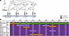 Figure 1
Discovery cohort study design and associations of biomarkers with fibrosis-like abnormalities. (A) Cohort study design of hospitalized survivors of severe and critical COVID-19, with sampling weighted for 50% invasive mechanical ventilation survivors. Schema of measurements and associations of blood biomarkers and high-resolution chest CT scans with sample size available at each time point. (B) Associations of serum biomarkers with fibrosis-like abnormalities. P values represent the P-for-association between the plasma biomarker and predictive risk of fibrotic pattern on thoracic CT using generalized additive models with LOESS smoothers, with adjustment for age, sex, race/ethnicity, BMI, COPD, asthma, pack-years history of smoking, estimated glomerular filtration rate, use of corticosteroids during COVID-19 hospitalization, IL-6 receptor inhibitor therapy during COVID-19 hospitalization, ventilator days, and days since initial SARS-CoV-2 infection utilizing covariate balanced propensity scores.
The prevalence of fibrosis-like abnormalities at follow-up in the Columbia and UBC cohorts ranged from 57%–64%, and was 43% in the nested case-control cohort from McGill, with 16 participants with and 21 participants without fibrosis-like abnormalities, matching on demographics, comorbidities, and either hospital discharge, 4-month, or 15-month thoracic CT follow-up time. Fibrosis-like abnormalities were predominantly characterized by reticulations and traction bronchiectasis. We observed honeycombing in less than 5% of Columbia and UBC patients and in 4 (11%) McGill patients (Supplemental Tables 2 and 3).
In the Columbia cohort, of the 18 biomarkers tested, only CC16 had consistent significant associations with the predicted risk of fibrosis-like abnormalities in adjusted analyses over 3 years of follow-up (Figure 1B). Sensitivity analyses revealed potential model overfitting with high outliers after natural log transformation observed in 6 end-stage renal disease (ESRD) dialysis patients. Since CC16 is renally excreted (22, 23), we report CC16 analyses excluding Columbia participants with ESRD and excluded ESRD patients in validation cohorts. CC16 declined slightly over 3 years but remained consistently higher among those with fibrosis-like abnormalities (Figure 2A), including in exploratory longitudinal analyses (Supplemental Figure 2). In 9 cross-sectional and longitudinal adjusted associations tested between hospital discharge and 3-year follow-up, CC16 had consistent, large magnitude, direct, and mostly linear associations with the predicted risk of fibrosis-like abnormalities (Figure 2B). The adjusted odds of fibrosis-like abnormalities for every natural log fold increase in CC16 ranged from 5.0 (95% CI: 1.6–20.7) to 16 (95% CI: 4.1–111). Adjusted odds ratios (ORs) increased 2- to 7-fold across tertiles of CC16 (all P-for-trend < 0.02) (Supplemental Table 4). Higher CC16 had a moderate inverse correlation with lower percentage predicted diffusion capacity of carbon monoxide (DLCO) at 4 months and 3 years but had no statistically significant correlation with DLCO at 15 months (Supplemental Table 5). In post hoc unadjusted and adjusted cross-sectional analyses, CC16 did not demonstrate consistent inverse associations with telomere length, with an association only detected between CC16 and telomere length modeled as a continuous variable at 4-month follow-up (Supplemental Table 6).
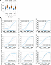 Figure 2
Associations of CC16 with fibrosis-like abnormalities in the discovery cohort. (A) Dot-box plots of plasma CC16 at hospital discharge, 4 months, 15 months, and 3 years, stratified by fibrotic pattern status in the Columbia (discovery) cohort. P values are derived from Mann-Whitney tests. Boxes represent the IQR, and the middle bars represent the median. (B) Plots of generalized additive models (GAM) with LOESS smoothers with adjustment for age, sex, race/ethnicity, BMI, COPD, asthma, pack-year history of smoking, estimated glomerular filtration rate, use of corticosteroids during COVID-19 hospitalization, IL-6 receptor inhibitor therapy during COVID-19 hospitalization, ventilator days, and days since initial SARS-CoV-2 infection using covariate balanced propensity scores.
In the UBC cohort, 3-month cross-sectional analyses revealed a direct, linear, borderline significant association between CC16 and the predicted risk of fibrosis-like abnormalities (generalized additive model [GAM] P = 0.058) that was statistically significant in unadjusted (P = 0.037) and adjusted logistic regression analyses (OR 2.92, 95% CI per natural log fold increase in CC16: 1.07–9.07) (Figure 3 and Supplemental Table 7). Higher CC16 had a moderate inverse correlation with lower percentage predicted DLCO (Supplemental Table 5).
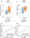 Figure 3
Associations of CC16 with fibrosis-like abnormalities in the validation cohorts. (A and C) Dox-box plots of plasma CC16 stratified by fibrotic pattern status in the University of British Columbia (UBC) and McGill University validation cohorts. Boxes represent the IQR and the middle bars represent the median. P values are derived from Mann-Whitney tests. (B and D) Plots of generalized additive models (GAM) with LOESS smoothers with adjustment for age, sex, race/ethnicity, BMI, COPD, asthma, pack-year history of smoking, estimated glomerular filtration rate, use of corticosteroids during COVID-19 hospitalization, IL-6 receptor inhibitor therapy during COVID-19 hospitalization, ventilator days, and days since initial SARS-CoV-2 infection using covariate balanced propensity scores.
Given the small sample size of the McGill cohort, we tested associations and estimated effect sizes of CC16 with fibrosis-like abnormalities for the hospital discharge, 4-month, and 15-month follow-up participants combined. We observed a similar direct, linear, but non-significant association in unadjusted and adjusted analyses (Figure 3, C and D), with an adjusted OR of fibrosis-like abnormalities of 2.36 (95% CI: 0.93–7.03) per natural log fold increase in CC16 (Supplemental Table 7). In unadjusted analyses stratified by follow-up time period, we observed nonsignificant higher median CC16 levels in those with fibrosis-like abnormalities (Supplemental Figure 3). In post hoc sensitivity analyses excluding 3 patients with possible honeycombing found on thoracic CT greater than 5 years prior to the COVID-19 pandemic, we observed the magnitude of the associations to be slightly less, with the data still collectively suggesting an association of higher CC16 levels with fibrosis-like abnormalities, even though the sample size becomes small in analyses stratified by follow-up time period (Supplemental Table 7 and Supplemental Figure 4).
In radiographic analyses of the Columbia cohort, higher levels of CC16 were directly and linearly associated with a higher airway-to-lung ratio at 15 months in adjusted longitudinal and cross-sectional analyses (Figure 4), with consistent effect sizes over time. For example, every natural log fold increase in CC16 at hospital discharge was associated with a 0.61 (0.21–1.01) standard deviation (SD) unit increase airway-to-lung ratio (Supplemental Table 8). Associations were robust to complete-case and larger airway (presubsegmental) sensitivity analyses (Supplemental Figure 5).
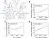 Figure 4
Associations of CC16 with airway-to-lung ratio measured on 15-month thoracic CT in the discovery cohort. (A) Using Apollo software (VIDA Diagnostics), airway-to-lung ratio was quantified on inspiratory thoracic CT performed at 15 months as the geometric mean of airway lumen diameters measured at 19 standard anatomic locations divided by the cube root of lung volume. (B–D) Plots are of generalized additive models (GAM) with LOESS smoothers with adjustment for age, sex, race/ethnicity, BMI, COPD, asthma, pack-year history of smoking, estimated glomerular filtration rate, use of corticosteroids during COVID-19 hospitalization, IL-6 receptor inhibitor therapy during COVID-19 hospitalization, ventilator days, and days since initial SARS-CoV-2 infection using covariate balanced propensity scores.
Functional analyses with lung tissue. The demographics and clinical characteristics of 4 COVID-19 survivors with the highest fibrosis scores on thoracic imaging at 3-year follow-up (24) and 4 control participants (lung transplant recipients without histopathologic evidence of rejection or infection, Supplemental Figure 6) are described in Supplemental Table 9 (Figure 5A). A single-cell suspension of each transbronchial lung biopsy sample was sorted by fluorescence-activated cell sorting (FACS) to mix epithelial, immune/endothelial, and non-epithelial/non-immune/non-endothelial cells in a 1:1:1 ratio prior to sequencing (Figure 5A). Clustering and distinction of epithelial cell types by the expression of top genes shows 2 groups of cells expressing SCGB1A1 (Figure 5, C–D). The first, designated as SCGB1A1+MUC5B+ airway cells, do not express surfactant genes but robustly express mucin genes; these are secretory cells (Figure 5E). The second, designated as SCGB1A1+SCGB3A2+SFTPB+ airway cells, are similar to the previously described preterminal bronchiole secretory cells (pre-TB-SC) or respiratory airways secretory (RAS) cells (Figure 5E and Supplemental Figure 7) (25, 26). There is evidence of increased proportions of both of these SCGB1A1-expressing cell clusters when analyzed separately (Figure 5F) or in combination (Figure 5G) in COVID-19 samples as compared with control samples. Proportions of SCGB1A1-expressing cell clusters were more dissimilar between COVID-19 and control samples than the proportions of alveolar type 1 and type 2 (AT1 and AT2) epithelial cells (Figure 5H), suggesting that post-COVID-19 effects drive expansion of airway rather than alveolar cells. Differences in fibroblast, immune, and endothelial populations between COVID-19 and control samples are shown in Supplemental Figure 7. None of the COVID-19 cases carried the MUC5B risk allele that has been linked to pulmonary fibrosis.
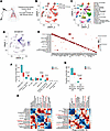 Figure 5
Increased proportions of SCGB1A1-expressing lung cells after severe COVID-19. (A) Schematic. (B) UMAP plot of cells from participants with control lung (aqua) and post–COVID-19 with fibrosis (red). (C) UMAP plot of annotated cell types. (D) Feature plot of SCGB1A1 expression. (E) Dot blot plot of top expression genes in each of the different cell clusters. (F) Proportion of different subsets of lung epithelial (EpCAM+) cells found in control and COVID-19 with fibrosis. (G) Proportion of SCGB1A1-expressing cells (from the SCGB1A1+MUC5B+ airway and SCGB1A1+SCGB3A2+SFTPB+ airway cell clusters) and non–SCGB1A1-expressing cells (all other EpCAM+ cell clusters) found in controls and COVID-19 with fibrosis. (H) Proportions of SCGB1A1-expressing cells and other cell types in controls and COVID-19 with fibrosis samples. P values are from Mann-Whitney (F) or Fischer’s exact tests (G).
We compared lung explants from 7 individuals who underwent lung transplantation 2–29 months after COVID-19 (Supplemental Table 10) to 11 normal control samples (lung resected adjacent to a pulmonary nodule) (Figure 6A). There was no difference between SCGB1A1/CC16 immunofluorescent staining of epithelial cells lining large (>100 μm) airways, but the post–COVID-19 lung explants showed dramatically increased expression in low cuboidal epithelium lining small (<100 μm) airways (Figure 6, B–D). Compared with control samples, COVID-19 explant samples (Figure 6E) had a greater than 3-fold increase in CC16 and MUC5B coexpression (0%–14% vs. 18%–26% cells, respectively; P = 0.0006) (Figure 6, F and G). These double-positive cells were typically located in small airways in areas of peribronchial metaplasia, with MUC5B staining the cell apices and extracellular spaces, and CC16 staining the basolateral portion of the cells. Photomicrographs of immunofluorescence antibody controls are shown in Supplemental Figure 8.
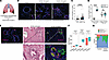 Figure 6
Increased SCGB1A1 immunofluorescence with MUC5B in small airways of the lung after severe COVID-19. (A) Schematic. (B) Immunofluorescence pattern of SCGB1A1 in large and small airways in control and COVID-19 explants (scale bars: 200 μm). Insets show staining at higher magnification (scale bars: 50 μm). (C) Automated quantification of SCGB1A1 immunofluorescence in control (n = 11) and COVID-19 explanted lungs (n = 7). Each point represents the average SCGB1A1+ areas divided by number of DAPI+ cells of 42 photomicrographs for each case at ×20 magnification. ****P < 2.2 × 10⁻¹⁶. (D) Manual counting of SCGB1A1+ airway epithelial cells in airways measuring less than or greater than 100 μm in diameter in 4 slides from all n = 7 COVID-19 explants and n = 7 sex-matched controls. ****P < 1 × 10⁻⁶ for small (<100 μm) airways. P = 0.67 for large airways. (E) Representative coimmunofluorescent staining of SCGB1A1 and MUC5B in control and COVID-19 explant tissue (scale bars: 100 μm). H&E staining of an adjacent cut from the COVID-19 explant is shown for reference. Higher-power magnification images of the H&E-stained slide and the immunofluorescence image from the areas denoted by the boxes are shown below. MUC5B signals (green) are seen not only in cells but also within the adjacent extracellular space. (F) Quantification of the absolute number of MUC5B+ cells, SCGB1A1+ cells, and MUC5B+SCGB1A1+ double-positive cells in COVID-19 explants (n = 7) and control lung tissue (n = 7). (G) Bar graph demonstrating the percentage of airway cells with the indicated staining pattern found in COVID-19 explants (n = 7) and control lung tissue (n = 7). P values are from Mann-Whitney tests.


