
Research ArticleGeneticsOncology
Open Access |  10.1172/jci.insight.192686
A tailored in vivo CRISPR screen identifies BAP1 as a potent tumor suppressor of sarcoma
Jianguo Huang,1 Xingliang Liu,1 Warren Floyd,2 William Haugh,1 Zhaoyu Sun,1 Melissa J. Kasiewicz,1 Yaping Wu,1 Brian Piening,1 John T. Welle,1 Wesley K. Rosales,1 Venkatesh Rajamanickam,1 So Young Kim,3 Eric S. Xu,2 Lixia Luo,2 Yan Ma,2 Rutulkumar Patel,2 Ziqiang Zhang,4 Brady Bernard,1 William L. Redmond,1 Walter J. Urba,1 R. Bryan Bell,1 and David G. Kirsch2,5,6,7,8
1Earle A. Chiles Research Institute, Providence Cancer Institute, Portland, Oregon, USA.
2Department of Radiation Oncology, Duke University Medical Center, Durham, North Carolina, USA.
3Department of Molecular Genetics and Microbiology, Duke University, Durham, North Carolina, USA.
4Department of Respiratory and Critical Care Medicine, Shanghai Pudong Hospital, Fudan University Pudong Medical Center, Pudong Hospital of Fudan University, Shanghai, China.
5Department of Pharmacology and Cancer Biology, Duke University Medical Center, Durham, North Carolina, USA.
6Radiation Medicine Program, Princess Margaret Cancer Center, University Health Network, Toronto, Ontario, Canada.
7Department of Radiation Oncology and
8Department of Medical Biophysics, University of Toronto, Toronto, Ontario, Canada.
Address correspondence to: Jianguo Huang, Earle A. Chiles Research Institute, Providence Cancer Institute, 4805 NE Glisan St., Suite 2N86, Portland, Oregon 97213, USA. Email: Jianguo.huang@providence.org. Or to: David G. Kirsch, Department of Radiation Oncology, Radiation Medicine Program, University of Toronto, Princess Margaret Cancer Center, 610 University Avenue, Toronto, Ontario M5G 2M9, Canada. Email: david.kirsch@uhn.ca.
Authorship note: JH and DGK contributed equally to this work and are co–corresponding authors.
Find articles by
Huang, J.
in:
PubMed
|
Google Scholar
|

1Earle A. Chiles Research Institute, Providence Cancer Institute, Portland, Oregon, USA.
2Department of Radiation Oncology, Duke University Medical Center, Durham, North Carolina, USA.
3Department of Molecular Genetics and Microbiology, Duke University, Durham, North Carolina, USA.
4Department of Respiratory and Critical Care Medicine, Shanghai Pudong Hospital, Fudan University Pudong Medical Center, Pudong Hospital of Fudan University, Shanghai, China.
5Department of Pharmacology and Cancer Biology, Duke University Medical Center, Durham, North Carolina, USA.
6Radiation Medicine Program, Princess Margaret Cancer Center, University Health Network, Toronto, Ontario, Canada.
7Department of Radiation Oncology and
8Department of Medical Biophysics, University of Toronto, Toronto, Ontario, Canada.
Address correspondence to: Jianguo Huang, Earle A. Chiles Research Institute, Providence Cancer Institute, 4805 NE Glisan St., Suite 2N86, Portland, Oregon 97213, USA. Email: Jianguo.huang@providence.org. Or to: David G. Kirsch, Department of Radiation Oncology, Radiation Medicine Program, University of Toronto, Princess Margaret Cancer Center, 610 University Avenue, Toronto, Ontario M5G 2M9, Canada. Email: david.kirsch@uhn.ca.
Authorship note: JH and DGK contributed equally to this work and are co–corresponding authors.
Find articles by Liu, X. in: PubMed | Google Scholar
1Earle A. Chiles Research Institute, Providence Cancer Institute, Portland, Oregon, USA.
2Department of Radiation Oncology, Duke University Medical Center, Durham, North Carolina, USA.
3Department of Molecular Genetics and Microbiology, Duke University, Durham, North Carolina, USA.
4Department of Respiratory and Critical Care Medicine, Shanghai Pudong Hospital, Fudan University Pudong Medical Center, Pudong Hospital of Fudan University, Shanghai, China.
5Department of Pharmacology and Cancer Biology, Duke University Medical Center, Durham, North Carolina, USA.
6Radiation Medicine Program, Princess Margaret Cancer Center, University Health Network, Toronto, Ontario, Canada.
7Department of Radiation Oncology and
8Department of Medical Biophysics, University of Toronto, Toronto, Ontario, Canada.
Address correspondence to: Jianguo Huang, Earle A. Chiles Research Institute, Providence Cancer Institute, 4805 NE Glisan St., Suite 2N86, Portland, Oregon 97213, USA. Email: Jianguo.huang@providence.org. Or to: David G. Kirsch, Department of Radiation Oncology, Radiation Medicine Program, University of Toronto, Princess Margaret Cancer Center, 610 University Avenue, Toronto, Ontario M5G 2M9, Canada. Email: david.kirsch@uhn.ca.
Authorship note: JH and DGK contributed equally to this work and are co–corresponding authors.
Find articles by Floyd, W. in: PubMed | Google Scholar
1Earle A. Chiles Research Institute, Providence Cancer Institute, Portland, Oregon, USA.
2Department of Radiation Oncology, Duke University Medical Center, Durham, North Carolina, USA.
3Department of Molecular Genetics and Microbiology, Duke University, Durham, North Carolina, USA.
4Department of Respiratory and Critical Care Medicine, Shanghai Pudong Hospital, Fudan University Pudong Medical Center, Pudong Hospital of Fudan University, Shanghai, China.
5Department of Pharmacology and Cancer Biology, Duke University Medical Center, Durham, North Carolina, USA.
6Radiation Medicine Program, Princess Margaret Cancer Center, University Health Network, Toronto, Ontario, Canada.
7Department of Radiation Oncology and
8Department of Medical Biophysics, University of Toronto, Toronto, Ontario, Canada.
Address correspondence to: Jianguo Huang, Earle A. Chiles Research Institute, Providence Cancer Institute, 4805 NE Glisan St., Suite 2N86, Portland, Oregon 97213, USA. Email: Jianguo.huang@providence.org. Or to: David G. Kirsch, Department of Radiation Oncology, Radiation Medicine Program, University of Toronto, Princess Margaret Cancer Center, 610 University Avenue, Toronto, Ontario M5G 2M9, Canada. Email: david.kirsch@uhn.ca.
Authorship note: JH and DGK contributed equally to this work and are co–corresponding authors.
Find articles by Haugh, W. in: PubMed | Google Scholar
1Earle A. Chiles Research Institute, Providence Cancer Institute, Portland, Oregon, USA.
2Department of Radiation Oncology, Duke University Medical Center, Durham, North Carolina, USA.
3Department of Molecular Genetics and Microbiology, Duke University, Durham, North Carolina, USA.
4Department of Respiratory and Critical Care Medicine, Shanghai Pudong Hospital, Fudan University Pudong Medical Center, Pudong Hospital of Fudan University, Shanghai, China.
5Department of Pharmacology and Cancer Biology, Duke University Medical Center, Durham, North Carolina, USA.
6Radiation Medicine Program, Princess Margaret Cancer Center, University Health Network, Toronto, Ontario, Canada.
7Department of Radiation Oncology and
8Department of Medical Biophysics, University of Toronto, Toronto, Ontario, Canada.
Address correspondence to: Jianguo Huang, Earle A. Chiles Research Institute, Providence Cancer Institute, 4805 NE Glisan St., Suite 2N86, Portland, Oregon 97213, USA. Email: Jianguo.huang@providence.org. Or to: David G. Kirsch, Department of Radiation Oncology, Radiation Medicine Program, University of Toronto, Princess Margaret Cancer Center, 610 University Avenue, Toronto, Ontario M5G 2M9, Canada. Email: david.kirsch@uhn.ca.
Authorship note: JH and DGK contributed equally to this work and are co–corresponding authors.
Find articles by Sun, Z. in: PubMed | Google Scholar
1Earle A. Chiles Research Institute, Providence Cancer Institute, Portland, Oregon, USA.
2Department of Radiation Oncology, Duke University Medical Center, Durham, North Carolina, USA.
3Department of Molecular Genetics and Microbiology, Duke University, Durham, North Carolina, USA.
4Department of Respiratory and Critical Care Medicine, Shanghai Pudong Hospital, Fudan University Pudong Medical Center, Pudong Hospital of Fudan University, Shanghai, China.
5Department of Pharmacology and Cancer Biology, Duke University Medical Center, Durham, North Carolina, USA.
6Radiation Medicine Program, Princess Margaret Cancer Center, University Health Network, Toronto, Ontario, Canada.
7Department of Radiation Oncology and
8Department of Medical Biophysics, University of Toronto, Toronto, Ontario, Canada.
Address correspondence to: Jianguo Huang, Earle A. Chiles Research Institute, Providence Cancer Institute, 4805 NE Glisan St., Suite 2N86, Portland, Oregon 97213, USA. Email: Jianguo.huang@providence.org. Or to: David G. Kirsch, Department of Radiation Oncology, Radiation Medicine Program, University of Toronto, Princess Margaret Cancer Center, 610 University Avenue, Toronto, Ontario M5G 2M9, Canada. Email: david.kirsch@uhn.ca.
Authorship note: JH and DGK contributed equally to this work and are co–corresponding authors.
Find articles by Kasiewicz, M. in: PubMed | Google Scholar
1Earle A. Chiles Research Institute, Providence Cancer Institute, Portland, Oregon, USA.
2Department of Radiation Oncology, Duke University Medical Center, Durham, North Carolina, USA.
3Department of Molecular Genetics and Microbiology, Duke University, Durham, North Carolina, USA.
4Department of Respiratory and Critical Care Medicine, Shanghai Pudong Hospital, Fudan University Pudong Medical Center, Pudong Hospital of Fudan University, Shanghai, China.
5Department of Pharmacology and Cancer Biology, Duke University Medical Center, Durham, North Carolina, USA.
6Radiation Medicine Program, Princess Margaret Cancer Center, University Health Network, Toronto, Ontario, Canada.
7Department of Radiation Oncology and
8Department of Medical Biophysics, University of Toronto, Toronto, Ontario, Canada.
Address correspondence to: Jianguo Huang, Earle A. Chiles Research Institute, Providence Cancer Institute, 4805 NE Glisan St., Suite 2N86, Portland, Oregon 97213, USA. Email: Jianguo.huang@providence.org. Or to: David G. Kirsch, Department of Radiation Oncology, Radiation Medicine Program, University of Toronto, Princess Margaret Cancer Center, 610 University Avenue, Toronto, Ontario M5G 2M9, Canada. Email: david.kirsch@uhn.ca.
Authorship note: JH and DGK contributed equally to this work and are co–corresponding authors.
Find articles by Wu, Y. in: PubMed | Google Scholar
1Earle A. Chiles Research Institute, Providence Cancer Institute, Portland, Oregon, USA.
2Department of Radiation Oncology, Duke University Medical Center, Durham, North Carolina, USA.
3Department of Molecular Genetics and Microbiology, Duke University, Durham, North Carolina, USA.
4Department of Respiratory and Critical Care Medicine, Shanghai Pudong Hospital, Fudan University Pudong Medical Center, Pudong Hospital of Fudan University, Shanghai, China.
5Department of Pharmacology and Cancer Biology, Duke University Medical Center, Durham, North Carolina, USA.
6Radiation Medicine Program, Princess Margaret Cancer Center, University Health Network, Toronto, Ontario, Canada.
7Department of Radiation Oncology and
8Department of Medical Biophysics, University of Toronto, Toronto, Ontario, Canada.
Address correspondence to: Jianguo Huang, Earle A. Chiles Research Institute, Providence Cancer Institute, 4805 NE Glisan St., Suite 2N86, Portland, Oregon 97213, USA. Email: Jianguo.huang@providence.org. Or to: David G. Kirsch, Department of Radiation Oncology, Radiation Medicine Program, University of Toronto, Princess Margaret Cancer Center, 610 University Avenue, Toronto, Ontario M5G 2M9, Canada. Email: david.kirsch@uhn.ca.
Authorship note: JH and DGK contributed equally to this work and are co–corresponding authors.
Find articles by Piening, B. in: PubMed | Google Scholar
1Earle A. Chiles Research Institute, Providence Cancer Institute, Portland, Oregon, USA.
2Department of Radiation Oncology, Duke University Medical Center, Durham, North Carolina, USA.
3Department of Molecular Genetics and Microbiology, Duke University, Durham, North Carolina, USA.
4Department of Respiratory and Critical Care Medicine, Shanghai Pudong Hospital, Fudan University Pudong Medical Center, Pudong Hospital of Fudan University, Shanghai, China.
5Department of Pharmacology and Cancer Biology, Duke University Medical Center, Durham, North Carolina, USA.
6Radiation Medicine Program, Princess Margaret Cancer Center, University Health Network, Toronto, Ontario, Canada.
7Department of Radiation Oncology and
8Department of Medical Biophysics, University of Toronto, Toronto, Ontario, Canada.
Address correspondence to: Jianguo Huang, Earle A. Chiles Research Institute, Providence Cancer Institute, 4805 NE Glisan St., Suite 2N86, Portland, Oregon 97213, USA. Email: Jianguo.huang@providence.org. Or to: David G. Kirsch, Department of Radiation Oncology, Radiation Medicine Program, University of Toronto, Princess Margaret Cancer Center, 610 University Avenue, Toronto, Ontario M5G 2M9, Canada. Email: david.kirsch@uhn.ca.
Authorship note: JH and DGK contributed equally to this work and are co–corresponding authors.
Find articles by Welle, J. in: PubMed | Google Scholar
1Earle A. Chiles Research Institute, Providence Cancer Institute, Portland, Oregon, USA.
2Department of Radiation Oncology, Duke University Medical Center, Durham, North Carolina, USA.
3Department of Molecular Genetics and Microbiology, Duke University, Durham, North Carolina, USA.
4Department of Respiratory and Critical Care Medicine, Shanghai Pudong Hospital, Fudan University Pudong Medical Center, Pudong Hospital of Fudan University, Shanghai, China.
5Department of Pharmacology and Cancer Biology, Duke University Medical Center, Durham, North Carolina, USA.
6Radiation Medicine Program, Princess Margaret Cancer Center, University Health Network, Toronto, Ontario, Canada.
7Department of Radiation Oncology and
8Department of Medical Biophysics, University of Toronto, Toronto, Ontario, Canada.
Address correspondence to: Jianguo Huang, Earle A. Chiles Research Institute, Providence Cancer Institute, 4805 NE Glisan St., Suite 2N86, Portland, Oregon 97213, USA. Email: Jianguo.huang@providence.org. Or to: David G. Kirsch, Department of Radiation Oncology, Radiation Medicine Program, University of Toronto, Princess Margaret Cancer Center, 610 University Avenue, Toronto, Ontario M5G 2M9, Canada. Email: david.kirsch@uhn.ca.
Authorship note: JH and DGK contributed equally to this work and are co–corresponding authors.
Find articles by Rosales, W. in: PubMed | Google Scholar
1Earle A. Chiles Research Institute, Providence Cancer Institute, Portland, Oregon, USA.
2Department of Radiation Oncology, Duke University Medical Center, Durham, North Carolina, USA.
3Department of Molecular Genetics and Microbiology, Duke University, Durham, North Carolina, USA.
4Department of Respiratory and Critical Care Medicine, Shanghai Pudong Hospital, Fudan University Pudong Medical Center, Pudong Hospital of Fudan University, Shanghai, China.
5Department of Pharmacology and Cancer Biology, Duke University Medical Center, Durham, North Carolina, USA.
6Radiation Medicine Program, Princess Margaret Cancer Center, University Health Network, Toronto, Ontario, Canada.
7Department of Radiation Oncology and
8Department of Medical Biophysics, University of Toronto, Toronto, Ontario, Canada.
Address correspondence to: Jianguo Huang, Earle A. Chiles Research Institute, Providence Cancer Institute, 4805 NE Glisan St., Suite 2N86, Portland, Oregon 97213, USA. Email: Jianguo.huang@providence.org. Or to: David G. Kirsch, Department of Radiation Oncology, Radiation Medicine Program, University of Toronto, Princess Margaret Cancer Center, 610 University Avenue, Toronto, Ontario M5G 2M9, Canada. Email: david.kirsch@uhn.ca.
Authorship note: JH and DGK contributed equally to this work and are co–corresponding authors.
Find articles by
Rajamanickam, V.
in:
PubMed
|
Google Scholar
|
1Earle A. Chiles Research Institute, Providence Cancer Institute, Portland, Oregon, USA.
2Department of Radiation Oncology, Duke University Medical Center, Durham, North Carolina, USA.
3Department of Molecular Genetics and Microbiology, Duke University, Durham, North Carolina, USA.
4Department of Respiratory and Critical Care Medicine, Shanghai Pudong Hospital, Fudan University Pudong Medical Center, Pudong Hospital of Fudan University, Shanghai, China.
5Department of Pharmacology and Cancer Biology, Duke University Medical Center, Durham, North Carolina, USA.
6Radiation Medicine Program, Princess Margaret Cancer Center, University Health Network, Toronto, Ontario, Canada.
7Department of Radiation Oncology and
8Department of Medical Biophysics, University of Toronto, Toronto, Ontario, Canada.
Address correspondence to: Jianguo Huang, Earle A. Chiles Research Institute, Providence Cancer Institute, 4805 NE Glisan St., Suite 2N86, Portland, Oregon 97213, USA. Email: Jianguo.huang@providence.org. Or to: David G. Kirsch, Department of Radiation Oncology, Radiation Medicine Program, University of Toronto, Princess Margaret Cancer Center, 610 University Avenue, Toronto, Ontario M5G 2M9, Canada. Email: david.kirsch@uhn.ca.
Authorship note: JH and DGK contributed equally to this work and are co–corresponding authors.
Find articles by Kim, S. in: PubMed | Google Scholar
1Earle A. Chiles Research Institute, Providence Cancer Institute, Portland, Oregon, USA.
2Department of Radiation Oncology, Duke University Medical Center, Durham, North Carolina, USA.
3Department of Molecular Genetics and Microbiology, Duke University, Durham, North Carolina, USA.
4Department of Respiratory and Critical Care Medicine, Shanghai Pudong Hospital, Fudan University Pudong Medical Center, Pudong Hospital of Fudan University, Shanghai, China.
5Department of Pharmacology and Cancer Biology, Duke University Medical Center, Durham, North Carolina, USA.
6Radiation Medicine Program, Princess Margaret Cancer Center, University Health Network, Toronto, Ontario, Canada.
7Department of Radiation Oncology and
8Department of Medical Biophysics, University of Toronto, Toronto, Ontario, Canada.
Address correspondence to: Jianguo Huang, Earle A. Chiles Research Institute, Providence Cancer Institute, 4805 NE Glisan St., Suite 2N86, Portland, Oregon 97213, USA. Email: Jianguo.huang@providence.org. Or to: David G. Kirsch, Department of Radiation Oncology, Radiation Medicine Program, University of Toronto, Princess Margaret Cancer Center, 610 University Avenue, Toronto, Ontario M5G 2M9, Canada. Email: david.kirsch@uhn.ca.
Authorship note: JH and DGK contributed equally to this work and are co–corresponding authors.
Find articles by
Xu, E.
in:
PubMed
|
Google Scholar
|
1Earle A. Chiles Research Institute, Providence Cancer Institute, Portland, Oregon, USA.
2Department of Radiation Oncology, Duke University Medical Center, Durham, North Carolina, USA.
3Department of Molecular Genetics and Microbiology, Duke University, Durham, North Carolina, USA.
4Department of Respiratory and Critical Care Medicine, Shanghai Pudong Hospital, Fudan University Pudong Medical Center, Pudong Hospital of Fudan University, Shanghai, China.
5Department of Pharmacology and Cancer Biology, Duke University Medical Center, Durham, North Carolina, USA.
6Radiation Medicine Program, Princess Margaret Cancer Center, University Health Network, Toronto, Ontario, Canada.
7Department of Radiation Oncology and
8Department of Medical Biophysics, University of Toronto, Toronto, Ontario, Canada.
Address correspondence to: Jianguo Huang, Earle A. Chiles Research Institute, Providence Cancer Institute, 4805 NE Glisan St., Suite 2N86, Portland, Oregon 97213, USA. Email: Jianguo.huang@providence.org. Or to: David G. Kirsch, Department of Radiation Oncology, Radiation Medicine Program, University of Toronto, Princess Margaret Cancer Center, 610 University Avenue, Toronto, Ontario M5G 2M9, Canada. Email: david.kirsch@uhn.ca.
Authorship note: JH and DGK contributed equally to this work and are co–corresponding authors.
Find articles by Luo, L. in: PubMed | Google Scholar
1Earle A. Chiles Research Institute, Providence Cancer Institute, Portland, Oregon, USA.
2Department of Radiation Oncology, Duke University Medical Center, Durham, North Carolina, USA.
3Department of Molecular Genetics and Microbiology, Duke University, Durham, North Carolina, USA.
4Department of Respiratory and Critical Care Medicine, Shanghai Pudong Hospital, Fudan University Pudong Medical Center, Pudong Hospital of Fudan University, Shanghai, China.
5Department of Pharmacology and Cancer Biology, Duke University Medical Center, Durham, North Carolina, USA.
6Radiation Medicine Program, Princess Margaret Cancer Center, University Health Network, Toronto, Ontario, Canada.
7Department of Radiation Oncology and
8Department of Medical Biophysics, University of Toronto, Toronto, Ontario, Canada.
Address correspondence to: Jianguo Huang, Earle A. Chiles Research Institute, Providence Cancer Institute, 4805 NE Glisan St., Suite 2N86, Portland, Oregon 97213, USA. Email: Jianguo.huang@providence.org. Or to: David G. Kirsch, Department of Radiation Oncology, Radiation Medicine Program, University of Toronto, Princess Margaret Cancer Center, 610 University Avenue, Toronto, Ontario M5G 2M9, Canada. Email: david.kirsch@uhn.ca.
Authorship note: JH and DGK contributed equally to this work and are co–corresponding authors.
Find articles by Ma, Y. in: PubMed | Google Scholar
1Earle A. Chiles Research Institute, Providence Cancer Institute, Portland, Oregon, USA.
2Department of Radiation Oncology, Duke University Medical Center, Durham, North Carolina, USA.
3Department of Molecular Genetics and Microbiology, Duke University, Durham, North Carolina, USA.
4Department of Respiratory and Critical Care Medicine, Shanghai Pudong Hospital, Fudan University Pudong Medical Center, Pudong Hospital of Fudan University, Shanghai, China.
5Department of Pharmacology and Cancer Biology, Duke University Medical Center, Durham, North Carolina, USA.
6Radiation Medicine Program, Princess Margaret Cancer Center, University Health Network, Toronto, Ontario, Canada.
7Department of Radiation Oncology and
8Department of Medical Biophysics, University of Toronto, Toronto, Ontario, Canada.
Address correspondence to: Jianguo Huang, Earle A. Chiles Research Institute, Providence Cancer Institute, 4805 NE Glisan St., Suite 2N86, Portland, Oregon 97213, USA. Email: Jianguo.huang@providence.org. Or to: David G. Kirsch, Department of Radiation Oncology, Radiation Medicine Program, University of Toronto, Princess Margaret Cancer Center, 610 University Avenue, Toronto, Ontario M5G 2M9, Canada. Email: david.kirsch@uhn.ca.
Authorship note: JH and DGK contributed equally to this work and are co–corresponding authors.
Find articles by Patel, R. in: PubMed | Google Scholar
1Earle A. Chiles Research Institute, Providence Cancer Institute, Portland, Oregon, USA.
2Department of Radiation Oncology, Duke University Medical Center, Durham, North Carolina, USA.
3Department of Molecular Genetics and Microbiology, Duke University, Durham, North Carolina, USA.
4Department of Respiratory and Critical Care Medicine, Shanghai Pudong Hospital, Fudan University Pudong Medical Center, Pudong Hospital of Fudan University, Shanghai, China.
5Department of Pharmacology and Cancer Biology, Duke University Medical Center, Durham, North Carolina, USA.
6Radiation Medicine Program, Princess Margaret Cancer Center, University Health Network, Toronto, Ontario, Canada.
7Department of Radiation Oncology and
8Department of Medical Biophysics, University of Toronto, Toronto, Ontario, Canada.
Address correspondence to: Jianguo Huang, Earle A. Chiles Research Institute, Providence Cancer Institute, 4805 NE Glisan St., Suite 2N86, Portland, Oregon 97213, USA. Email: Jianguo.huang@providence.org. Or to: David G. Kirsch, Department of Radiation Oncology, Radiation Medicine Program, University of Toronto, Princess Margaret Cancer Center, 610 University Avenue, Toronto, Ontario M5G 2M9, Canada. Email: david.kirsch@uhn.ca.
Authorship note: JH and DGK contributed equally to this work and are co–corresponding authors.
Find articles by Zhang, Z. in: PubMed | Google Scholar
1Earle A. Chiles Research Institute, Providence Cancer Institute, Portland, Oregon, USA.
2Department of Radiation Oncology, Duke University Medical Center, Durham, North Carolina, USA.
3Department of Molecular Genetics and Microbiology, Duke University, Durham, North Carolina, USA.
4Department of Respiratory and Critical Care Medicine, Shanghai Pudong Hospital, Fudan University Pudong Medical Center, Pudong Hospital of Fudan University, Shanghai, China.
5Department of Pharmacology and Cancer Biology, Duke University Medical Center, Durham, North Carolina, USA.
6Radiation Medicine Program, Princess Margaret Cancer Center, University Health Network, Toronto, Ontario, Canada.
7Department of Radiation Oncology and
8Department of Medical Biophysics, University of Toronto, Toronto, Ontario, Canada.
Address correspondence to: Jianguo Huang, Earle A. Chiles Research Institute, Providence Cancer Institute, 4805 NE Glisan St., Suite 2N86, Portland, Oregon 97213, USA. Email: Jianguo.huang@providence.org. Or to: David G. Kirsch, Department of Radiation Oncology, Radiation Medicine Program, University of Toronto, Princess Margaret Cancer Center, 610 University Avenue, Toronto, Ontario M5G 2M9, Canada. Email: david.kirsch@uhn.ca.
Authorship note: JH and DGK contributed equally to this work and are co–corresponding authors.
Find articles by Bernard, B. in: PubMed | Google Scholar
1Earle A. Chiles Research Institute, Providence Cancer Institute, Portland, Oregon, USA.
2Department of Radiation Oncology, Duke University Medical Center, Durham, North Carolina, USA.
3Department of Molecular Genetics and Microbiology, Duke University, Durham, North Carolina, USA.
4Department of Respiratory and Critical Care Medicine, Shanghai Pudong Hospital, Fudan University Pudong Medical Center, Pudong Hospital of Fudan University, Shanghai, China.
5Department of Pharmacology and Cancer Biology, Duke University Medical Center, Durham, North Carolina, USA.
6Radiation Medicine Program, Princess Margaret Cancer Center, University Health Network, Toronto, Ontario, Canada.
7Department of Radiation Oncology and
8Department of Medical Biophysics, University of Toronto, Toronto, Ontario, Canada.
Address correspondence to: Jianguo Huang, Earle A. Chiles Research Institute, Providence Cancer Institute, 4805 NE Glisan St., Suite 2N86, Portland, Oregon 97213, USA. Email: Jianguo.huang@providence.org. Or to: David G. Kirsch, Department of Radiation Oncology, Radiation Medicine Program, University of Toronto, Princess Margaret Cancer Center, 610 University Avenue, Toronto, Ontario M5G 2M9, Canada. Email: david.kirsch@uhn.ca.
Authorship note: JH and DGK contributed equally to this work and are co–corresponding authors.
Find articles by
Redmond, W.
in:
PubMed
|
Google Scholar
|
1Earle A. Chiles Research Institute, Providence Cancer Institute, Portland, Oregon, USA.
2Department of Radiation Oncology, Duke University Medical Center, Durham, North Carolina, USA.
3Department of Molecular Genetics and Microbiology, Duke University, Durham, North Carolina, USA.
4Department of Respiratory and Critical Care Medicine, Shanghai Pudong Hospital, Fudan University Pudong Medical Center, Pudong Hospital of Fudan University, Shanghai, China.
5Department of Pharmacology and Cancer Biology, Duke University Medical Center, Durham, North Carolina, USA.
6Radiation Medicine Program, Princess Margaret Cancer Center, University Health Network, Toronto, Ontario, Canada.
7Department of Radiation Oncology and
8Department of Medical Biophysics, University of Toronto, Toronto, Ontario, Canada.
Address correspondence to: Jianguo Huang, Earle A. Chiles Research Institute, Providence Cancer Institute, 4805 NE Glisan St., Suite 2N86, Portland, Oregon 97213, USA. Email: Jianguo.huang@providence.org. Or to: David G. Kirsch, Department of Radiation Oncology, Radiation Medicine Program, University of Toronto, Princess Margaret Cancer Center, 610 University Avenue, Toronto, Ontario M5G 2M9, Canada. Email: david.kirsch@uhn.ca.
Authorship note: JH and DGK contributed equally to this work and are co–corresponding authors.
Find articles by Urba, W. in: PubMed | Google Scholar
1Earle A. Chiles Research Institute, Providence Cancer Institute, Portland, Oregon, USA.
2Department of Radiation Oncology, Duke University Medical Center, Durham, North Carolina, USA.
3Department of Molecular Genetics and Microbiology, Duke University, Durham, North Carolina, USA.
4Department of Respiratory and Critical Care Medicine, Shanghai Pudong Hospital, Fudan University Pudong Medical Center, Pudong Hospital of Fudan University, Shanghai, China.
5Department of Pharmacology and Cancer Biology, Duke University Medical Center, Durham, North Carolina, USA.
6Radiation Medicine Program, Princess Margaret Cancer Center, University Health Network, Toronto, Ontario, Canada.
7Department of Radiation Oncology and
8Department of Medical Biophysics, University of Toronto, Toronto, Ontario, Canada.
Address correspondence to: Jianguo Huang, Earle A. Chiles Research Institute, Providence Cancer Institute, 4805 NE Glisan St., Suite 2N86, Portland, Oregon 97213, USA. Email: Jianguo.huang@providence.org. Or to: David G. Kirsch, Department of Radiation Oncology, Radiation Medicine Program, University of Toronto, Princess Margaret Cancer Center, 610 University Avenue, Toronto, Ontario M5G 2M9, Canada. Email: david.kirsch@uhn.ca.
Authorship note: JH and DGK contributed equally to this work and are co–corresponding authors.
Find articles by
Bell, R.
in:
PubMed
|
Google Scholar
|
1Earle A. Chiles Research Institute, Providence Cancer Institute, Portland, Oregon, USA.
2Department of Radiation Oncology, Duke University Medical Center, Durham, North Carolina, USA.
3Department of Molecular Genetics and Microbiology, Duke University, Durham, North Carolina, USA.
4Department of Respiratory and Critical Care Medicine, Shanghai Pudong Hospital, Fudan University Pudong Medical Center, Pudong Hospital of Fudan University, Shanghai, China.
5Department of Pharmacology and Cancer Biology, Duke University Medical Center, Durham, North Carolina, USA.
6Radiation Medicine Program, Princess Margaret Cancer Center, University Health Network, Toronto, Ontario, Canada.
7Department of Radiation Oncology and
8Department of Medical Biophysics, University of Toronto, Toronto, Ontario, Canada.
Address correspondence to: Jianguo Huang, Earle A. Chiles Research Institute, Providence Cancer Institute, 4805 NE Glisan St., Suite 2N86, Portland, Oregon 97213, USA. Email: Jianguo.huang@providence.org. Or to: David G. Kirsch, Department of Radiation Oncology, Radiation Medicine Program, University of Toronto, Princess Margaret Cancer Center, 610 University Avenue, Toronto, Ontario M5G 2M9, Canada. Email: david.kirsch@uhn.ca.
Authorship note: JH and DGK contributed equally to this work and are co–corresponding authors.
Find articles by
Kirsch, D.
in:
PubMed
|
Google Scholar
|
Published June 30, 2026 - More info
JCI Insight. 2026;11(16):e192686. https://doi.org/10.1172/jci.insight.192686.
© 2026 Huang et al. This work is licensed under the Creative Commons Attribution 4.0 International License. To view a copy of this license, visit http://creativecommons.org/licenses/by/4.0/.
Received: February 20, 2025; Accepted: June 24, 2026


-
Results
Intramuscular injection of a CRISPR sgRNA library induces spontaneous sarcomas in mice. To investigate the tumor suppressor function of genes frequently altered in human UPS, we employed a direct in vivo autochthonous screening strategy. This approach enables targeted mutagenesis within the native skeletal muscle microenvironment of mice (38). Using data from The Cancer Genome Atlas (TCGA) and the Genomics Evidence Neoplasia Information Exchange (GENIE), we identified the top 35 genes with loss-of-function mutations or CNAs in human UPS and myxofibrosarcoma (MFS), which share similar genetic profiles (Supplemental Table 1; supplemental material available online with this article; https://doi.org/10.1172/jci.insight.192686DS1), along with tumor protein p53 (TP53) and cyclin-dependent kinase inhibitor 2A (CDKN2A) (39). We designed a custom single-guide RNA (sgRNA) library targeting these 35 genes (excluding Trp53 and Cdkn2a), with 4 sgRNAs per gene sourced from the mouse Brie library (Supplemental Table 2) (40). Additionally, we included sgRNAs targeting 5 housekeeping genes: ribosomal protein L7 (Rpl7), ribosomal protein 19 (Rps19), ribosomal protein L22 (Rpl22), ribosomal protein 18 (Rps18), and ribosomal protein 11 (Rps11) (38). The customized sgRNA library also included 8 nontargeting control sgRNAs from the mouse Brie library (Supplemental Table 2). Given that TP53 is mutated in approximately 65% of human UPSs and MFSs (39), and that deletion of Cdkn2a can substitute for Trp53 loss to induce sarcomagenesis in conjunction with KrasG12D (41), we cloned this customized sgRNA library into 3 different plasmid backbones: (a) negative control sgRNA–expressing plasmid (pX334–Negative sgRNA library); (b) Trp53 sgRNA–expressing plasmid (pX334–Trp53 sgRNA library); and (c) Cdkn2a sgRNA–expressing plasmid (pX334–Cdkn2a sgRNA library) (Figure 1A). The Trp53 sgRNA and Cdkn2a sgRNA in these constructs have been confirmed to induce robust and specific editing in the targeting loci, according to previous studies (41, 42). All 3 plasmid libraries were sequenced to confirm full coverages of all sgRNAs (Supplemental Figure 1). We used an in vivo electroporation (IVE) method to deliver these plasmids into the gastrocnemius muscle of Cas9-expressing mice, enabling CRISPR/Cas9–mediated mutagenesis of tumor suppressor genes and generation of UPS-like tumors (11, 41). Only the pX334–Trp53 sgRNA library induced tumors in both Rosa26loxP-Cas9/loxP-Cas9 and Rosa26loxP-Cas9/+ mice (Figure 1B and Supplemental Figure 2). In an independent cohort, we confirmed that the pX334–Trp53 sgRNA library induced tumors in Rosa26loxP-Cas9/loxP-Cas9 mice, whereas the pX334–Trp53 sgRNA alone (lacking sgRNAs for other tumor suppressors) failed to induce tumors (Figure 1C). These results suggest that mutation of at least one additional gene from the library is required to cooperate with Trp53 loss in driving tumorigenesis.
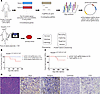 Figure 1
In vivo CRISPR/Cas9 screen identifies tumor suppressors of UPS in Rosa26loxP-Cas9 mice. (A) Schematic of the direct in vivo CRISPR/Cas9 screening strategy. (B) Two primary tumors developed in Rosa26loxP-Cas9/loxP-Cas9 mice following intramuscular delivery of the pX334–Trp53 sgRNA library (n = 5), while no tumors formed in mice receiving the pX334–Negative sgRNA library or pX334–Cdkn2a sgRNA library (n = 5 each). (C) In a separate cohort, 5 tumors developed in mice injected with the pX334–Trp53 sgRNA library (n = 13), but no tumors formed in mice injected with pX334–Trp53 sgRNA alone (n = 10). (D) Representative H&E and IHC staining (MyoD, myogenin, cytokeratin, S100) of a tumor induced by the pX334–Trp53 sgRNA library. Scale bars: 100 μm. For all experiments, n denotes biologically independent mice. Representative images are from experiments performed at least twice with similar results.
To characterize the tumors induced by the pX334–Trp53 sgRNA library, we performed hematoxylin and eosin (H&E) staining and immunohistochemistry (IHC) on sections from 10 tumors using antibodies against cytokeratin, MyoD, myogenin, and S100 (Figure 1D). A blinded pathological review determined that all tumors were poorly differentiated, negative for cytokeratin and S100, and weak or negative for MyoD and myogenin, consistent with UPS or myogenic UPS phenotypes.
Targeted-capture sequencing identifies multiple tumor suppressor candidates from the Trp53 library–induced tumors. We generated a total of 14 tumors in mice using the pX334–Trp53 sgRNA library alone, and an additional 3 tumors following co-delivery of the pX334–Trp53 sgRNA library with a Pten sgRNA (Supplemental Figure 3). To validate CRISPR/Cas9–mediated gene editing using our IVE method, we injected the pX334–Trp53 sgRNA library into the gastrocnemius muscle of 2 mice. Four weeks after injection, the muscle tissues were harvested, and genomic DNA was extracted from both tumors (S1–S17) and injected muscle tissues (S18–S19) for targeted-capture sequencing (38).
Bioinformatic analysis revealed CRISPR/Cas9–mediated editing in Cysltr2 (S18) and Pask (S19) in the injected muscle tissues. However, editing was not detected in most targeted genes, including Trp53, in these samples. This may be due to the absence of transformed cells harboring Trp53 mutations in the harvested tissues or the inability of our sequencing method to detect low-frequency edits in underrepresented mutant cells. Interestingly, CYSLTR2 ranks among the top 3 genes with loss-of-function mutations or CNAs in human UPS and MFS, following TP53 and RB1. PASK also shows the highest mutation frequency in UPS and MFS compared with other cancers in the TCGA database. These findings confirm that our IVE method can induce gene editing in vivo using pooled plasmid libraries (Figure 2A).
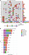 Figure 2
Targeted-capture sequencing of tumors induced by the pX334–Trp53 sgRNA library. (A) Mutation profiles from targeted-capture sequencing of injected muscle tissues (S18–S19) and tumors (S1–S17), including 3 tumors with additional Pten mutations (S9, S13, S14). (B) Top 10 most frequently mutated genes across all tumors.
In tumors generated by co-delivery of the pX334–Trp53 sgRNA library and the Pten sgRNA, Pten editing was detected exclusively in those samples, whereas no Pten edits were observed in tumors induced by the pX334–Trp53 sgRNA library alone (Figure 2A and Supplemental Figure 3). Editing of Trp53 was detected in nearly all tumors, although 4 tumors (S1, S7, S11, and S13) showed no detectable edits by targeted-capture sequencing. To further investigate, we performed T7 endonuclease I (T7E1) assays and Sanger sequencing on genomic DNA from these tumors. In tumor S1, we confirmed 82% insertions and deletions (indels) at the Trp53 locus, including a 3-nucleotide deletion in one allele (Supplemental Figure 4, A and B). It is possible that the other allele harbors a large deletion undetectable by our method, consistent with previous reports of large CRISPR-induced deletions in Trp53 (11). Owing to limitations in Sanger sequencing sensitivity (43), we performed next-generation sequencing on tumors S7, S11, and S13. We detected approximately 9% indels in S7, 11% in S11, and 12% in S13, with multiple indel variants per sample (Supplemental Table 3 and Supplemental Figure 4C). The relatively low indel percentages may reflect the presence of normal cells mixed with tumor tissue. Beyond Trp53, other frequently mutated genes in the tumors included Mllt3 (also known as Af9), Fat1, Bap1, and Rb1 (Figure 2, A and B). Notably, only Trp53 and Bap1 were mutated in tumor S3. According to TCGA data, BAP1 mutations or CNAs are mainly found in UPS among all STS subtypes (Figure 3A). Tumor S6 harbored mutations in Trp53 and Pask. To further investigate the role of these candidate tumor suppressors, we individually evaluated their ability to cooperate with Trp53 mutation in driving sarcoma development.
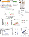 Figure 3
Validation of Bap1 as a potent tumor suppressor in UPS. (A) Mutation rates of 3 genes (TP53, RB1, and BAP1) in human UPS/MFS according to TCGA database. (B) IHC of a human sarcoma tissue array shows that BAP1 expression is significantly reduced in UPS, MFS, fibrosarcoma, and dermatofibrosarcoma compared with normal muscle (NM). Scale bars: 50 μm. Quantification was performed on independent tissue cores, and statistical significance was assessed using a 2-tailed Student’s t test. (C) IVE of plasmids expressing sgRNAs targeting Trp53 and Bap1 or Trp53 and Rb1 induced tumors in Rosa26loxP-Cas9/loxP-Cas9 mice. Tumor-free survival (latency to tumor formation) was analyzed using Kaplan-Meier curves and compared using the log-rank test. (D) Tumor quadrupling times were similar between PR CRISPR and PB CRISPR tumors. Statistical comparisons were performed using a 2-tailed Student’s t test. (E) Growth curves of representative PR and PB CRISPR tumors. (F) IVE of Cre recombinase and sgRNA targeting Bap1 (n = 11) induced significantly higher tumor penetrance compared with Cre alone or Cre plus sgRNA targeting Crebbp (3 tumors in 18 mice). Tumor-free survival (latency to tumor formation) was analyzed using Kaplan-Meier curves and compared using the log-rank test. (G) PRB Cre/CRISPR tumors grew significantly faster than PR Cre tumors. Statistical comparisons were performed using a 2-tailed Student’s t test. (H) Growth curves of representative PR Cre and PRB Cre/CRISPR tumors. For all experiments, n denotes biologically independent mice. Data are presented as mean ± SEM unless otherwise indicated. Representative images and growth curves are from experiments repeated at least twice with similar results.
Validation of tumor suppressor genes cooperating with Trp53 mutation to drive sarcoma development in mice. To evaluate BAP1 expression in human sarcomas, we performed IHC on normal skeletal muscle and a panel of sarcoma specimens, including undifferentiated pleomorphic sarcoma (UPS), myxofibrosarcoma (MFS), fibrosarcoma, and dermatofibrosarcoma, using tissue microarrays stained with BAP1 antibody (Figure 3B). BAP1 protein expression was significantly reduced in UPS, MFS, fibrosarcoma, and dermatofibrosarcoma compared with normal skeletal muscle. To validate candidate tumor suppressor genes identified from the CRISPR screen, we performed intramuscular injections of plasmids expressing sgRNAs targeting individual genes along with Trp53 sgRNA into Rosa26loxP-Cas9/loxP-Cas9 mice. Tumor formation was observed in mice electroporated with plasmids targeting Trp53 and Bap1, as well as those targeting Trp53 and Rb1 (Figure 3C). In contrast, no tumors were observed in mice injected with plasmids targeting Trp53 in combination with Cysltr2, Mst1r, Mllt3, Crebbp, Pbrm1, or Fat1 (Supplemental Figure 5). We next compared tumor growth rates between different genotypes. Tumors generated by deletion of Trp53 and Rb1 (referred to as PR CRISPR tumors) and those generated by deletion of Trp53 and Bap1 (PB CRISPR tumors) showed no significant difference in time to quadrupling of tumor volume (Figure 3, D and E).
To further explore genetic cooperation, we investigated whether additional mutations could accelerate tumorigenesis in the context of Trp53 and Rb1 loss. Previous studies in small cell lung cancer models have shown that Crebbp deletion accelerates tumor formation when combined with Trp53 and Rb1 loss (44). TCGA data also indicate that BAP1 copy number loss frequently co-occurs with RB1 loss in human UPS (Figure 3A). Therefore, we generated a transgenic mouse model of Trp53fl/fl Rb1fl/fl Rosa26loxP-Cas9/loxP-Cas9 (PR-loxP-Cas9 mice) and performed IVE to deliver plasmids expressing Cre recombinase and sgRNAs targeting either no gene, Bap1, or Crebbp. Targeting Crebbp did not affect tumor development initiated by Cre-mediated deletion of Trp53 and Rb1 (Figure 3F). In contrast, additional mutation of Bap1 significantly accelerated tumor onset and increased tumor penetrance in PR-loxP-Cas9 mice (Figure 3F). We then compared tumor growth rates between PR Cre tumors (deletion of Trp53 and Rb1) and PRB Cre/CRISPR tumors (deletion of Trp53, Rb1, and Bap1). PRB Cre/CRISPR tumors exhibited significantly faster growth, with shorter times to quadrupling of tumor volume (Figure 3, G and H). These findings suggest that Bap1 loss enhances sarcoma aggressiveness in the context of Trp53 and Rb1 deficiency.
Additionally, we observed lung metastases in mice bearing PR CRISPR tumors (1/11), PR Cre tumors (1/2), PB CRISPR tumors (4/16), and PRB Cre/CRISPR tumors (1/6). Multiple metastatic nodules were detected in the lungs of mice with PB CRISPR tumors (Supplemental Table 4). Histological analysis via H&E staining confirmed that all tumors were high-grade poorly differentiated sarcomas resembling UPS (Supplemental Figure 6).
Taken together, these results demonstrate that Bap1 functions as a potent tumor suppressor in UPS, both in the context of Trp53 loss alone and in combination with Rb1 loss.
High-throughput RNA sequencing reveals immune suppression in Bap1-loss mouse sarcomas. To systematically investigate the signaling pathways contributing to sarcoma development, we performed total RNA sequencing on tumors from 3 genotypes: PB CRISPR (n = 10), PR Cre (n = 5), and PRB Cre/CRISPR (n = 7), along with normal skeletal muscle controls. Bioinformatic analysis and gene set enrichment analysis (GSEA) revealed that PB CRISPR tumors were significantly enriched for oncogenic pathways, including the E2F targets, G2/M checkpoint, and MYC targets hallmarks, compared with normal muscle tissue (Figure 4, A and B) (10). Notably, multiple immune-related pathways were significantly downregulated in PB CRISPR tumors relative to normal muscle, including B cell receptor signaling pathways, B cell activation pathways, activation of immune response pathways, and major histocompatibility complex (MHC) class II protein complex assembly pathways (Figure 4, B and C). These findings suggest that Bap1 loss is associated with an immunosuppressive tumor microenvironment, consistent with previous observations in BAP1-driven uveal melanoma (45). Additionally, recent studies have shown that BAP1 loss leads to downregulation of MHC class II expression in B cell lymphoma (28). To further assess the similarity between PB CRISPR tumors and human UPS, we compared the hallmark pathways significantly regulated in PB CRISPR tumors versus normal muscle with those significantly regulated in human UPS versus normal muscle. This analysis revealed a significant correlation between PB CRISPR tumors and human UPS (Figure 4D; R = 0.36538, P = 0.00944), further supporting that PB CRISPR tumors recapitulate key molecular features of human UPS.
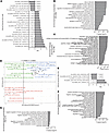 Figure 4
RNA sequencing reveals immune suppression in Bap1-deficient sarcomas. (A–C) PB CRISPR tumors show upregulation of E2F targets, G2/M checkpoint, MYC targets, DNA repair, and histone modification pathways and downregulation of immune response pathways versus normal muscle. (D) Comparison of hallmark pathways significantly dysregulated in human UPS (TCGA) versus normal human muscle and mouse PB CRISPR tumors versus normal mouse muscle revealed a significant correlation (R = 0.36538, P = 0.00944). Statistical significance was assessed using Pearson’s correlation analysis. (E–G) PRB Cre/CRISPR tumors exhibit upregulation of KRAS and MYC pathways and downregulation of B cell signaling and immune activation pathways versus PR Cre tumors. RNA-sequencing analyses were performed on biologically independent samples (PB CRISPR, n = 10; PR Cre, n = 5; PRB Cre/CRISPR, n = 7; normal muscle, n = 5). For GSEA, normalized enrichment scores and adjusted P values (FDR) were calculated as implemented in the fgsea package.
Given that Bap1 deletion increased sarcoma aggressiveness in the context of Trp53 and Rb1 loss (Figure 3D), we next compared PR Cre tumors with PRB Cre/CRISPR tumors. PRB Cre/CRISPR tumors exhibited significant upregulation of oncogenic pathways, including the hallmarks of KRAS signaling, unfolded protein response, and MYC targets (Figure 4E). These dysregulated pathways may contribute to the enhanced tumor penetrance and aggressiveness observed in PRB Cre/CRISPR tumors (46, 47). Furthermore, PRB Cre/CRISPR tumors showed significant downregulation of immune-related pathways compared with PR Cre tumors, including regulation of tumor necrosis factor (TNF) production, regulation of inflammatory response, and cytokine production in immune response (Figure 4, F and G). Collectively, these results indicate that Bap1 loss contributes to immune suppression in STSs, potentially facilitating tumor progression and resistance to immune surveillance.
Immune cell profiling suggests an immunosuppressive microenvironment in Bap1-loss mouse sarcomas. To examine the tumor immune microenvironment in mouse sarcomas driven by different genetic mutations, we performed multiplex IHC (48) and flow cytometry on primary tumors (49). Using a multiplex IHC panel including CD3, CD8, CD68, PD-1, CD20, and DAPI, we observed minimal infiltration of CD3+ T cells, CD8+ T cells, and CD20+ B cells in KP mouse sarcomas (driven by KrasG12D activation and Trp53 deletion in KrasLSL-G12D/+ Trp53fl/fl mice), consistent with previous reports (n = 1, data not shown) (50, 51). In contrast, PR CRISPR tumors (n = 4) exhibited significantly higher infiltration of CD3+ T cells, CD8+ T cells, and CD20+ B cells compared with PB CRISPR tumors (n = 5) (Figure 5A). To further validate the impact of Bap1 loss on immune cell infiltration, we compared PR Cre tumors (n = 3) with PRB Cre/CRISPR tumors (n = 4). PR Cre tumors showed significantly more CD3+ T cells, while CD8+ T cell and CD20+ B cell levels were comparable between groups (Figure 5B). CD68+ macrophages were abundant across all tumor types, consistent with findings in human UPS (Supplemental Figure 7) (52, 53).
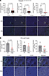 Figure 5
Multiplex IHC reveals reduced immune cell infiltration in Bap1-deficient sarcomas. (A) PR CRISPR tumors (n = 4) show significantly more CD3+ T cells than PB CRISPR tumors (n = 6). (B) PR Cre tumors (n = 3) show significantly more CD3+ T cells than PRB Cre/CRISPR tumors (n = 4). Each point represents an individual tumor. Scale bars: 100 μm. Statistical significance was assessed using a 2-tailed Student’s t test. For all experiments, n denotes biologically independent tumors. Data are presented as mean ± SEM. Representative images are from experiments performed at least twice with similar results.
We next used flow cytometry to analyze immune cell populations in tumors of varying sizes. We compared normal muscle (NM; n = 5), PR CRISPR large tumors (n = 4), PB CRISPR small tumors (<250 mm3; n = 3), and PB CRISPR large tumors (>1,000 mm3; n = 9) (Figure 6A). No significant differences were observed in CD8+ T cells among CD45+ cells. However, CD4+ T cell percentages were significantly lower in PB CRISPR large tumors compared with NM and PB early tumors. Notably, the ratio of CD4+ regulatory T cells (Tregs) to CD4+ T cells was significantly elevated in PB CRISPR large tumors. Further analysis revealed a higher frequency of proliferating (Ki-67+) CD8+ T cells in PR CRISPR large tumors compared with NM, PB large, and PB small tumors. Additionally, Ki-67+ FoxP3+ CD4+ Tregs were more abundant in PR CRISPR large tumors than in PB CRISPR large tumors. Although Ki-67+ Treg levels were lower in PB CRISPR large tumors than in PB small tumors, the difference was not statistically significant. PD-1 expression on CD8+ T cells did not differ significantly across tumor types (Supplemental Figure 8). These findings suggest that CD8+ T cells are more proliferative in PR CRISPR tumors than in PB CRISPR tumors.
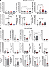 Figure 6
Flow cytometry analysis reveals immunosuppressive features in Bap1-deficient sarcomas. (A) No differences in CD8+ T cell percentages among CD45+ cells across NM (n = 5), PR CRISPR large (n = 5), PB CRISPR large (n = 9), and PB CRISPR small (n = 3) tumors. CD4+ T cell percentages were lower in PB CRISPR large tumors, with higher Treg/CD4+ ratios. PR CRISPR large tumors had more Ki-67+ CD8+ T cells and FoxP3– CD4+ effector T (Teff) cells. (B) No differences in CD8+ T cell, CD4+ T cell, or Treg percentages between PR Cre (n = 5) and PRB Cre/CRISPR (n = 3) tumors. PR Cre tumors had more Ki-67+ CD8+ T cells, Tim3+ CD8+ T cells, and CD11b+ cells. PRB Cre/CRISPR tumors had higher PMN-MDSC percentages and lower TAM percentages. PD-L1+ PMN-MDSCs were more abundant in PR Cre tumors. Each point represents an individual tumor or muscle. Statistical comparisons among multiple groups in A were performed using 1-way ANOVA followed by appropriate post hoc tests. Comparisons between 2 groups in B were performed using a 2-tailed Student’s t test. For all experiments, n denotes biologically independent samples. Data are presented as mean ± SEM.
We also profiled immune cells in PR Cre tumors (n = 5) and PRB Cre/CRISPR tumors (n = 3) (Figure 6B). PR Cre tumors exhibited significantly higher frequencies of proliferating (Ki-67+) CD8+ T cells and reduced expression of CD62L, a marker of T cell activation (54). Tim3+ CD8+ T cells were significantly lower in PR Cre tumors, while PD-1+ CD8+ T cell levels were similar between groups. These results suggest that CD8+ T cells in Bap1-wild-type tumors (PR Cre) are more proliferative and activated than those in Bap1-deficient tumors (PRB Cre/CRISPR).
We further examined myeloid-derived suppressor cells (MDSCs) and tumor-associated macrophages (TAMs). CD11b+ cell percentages among CD45+ cells were significantly higher in PB CRISPR large tumors compared with PB early tumors (Supplemental Figure 9). No differences were observed in CD11b+ cell percentages between PR Cre and PRB Cre/CRISPR tumors (Figure 6B). Additionally, there were no significant differences in polymorphonuclear MDSCs (PMN-MDSCs; CD11b+Ly6CintLy6Ghi), monocytic MDSCs (M-MDSCs; CD11b+Ly6ChiLy6Glo), or TAMs (CD11b+Ly6CloLy6GloF4/80+) between PB CRISPR large and small tumors (Supplemental Figure 9). Interestingly, PMN-MDSC percentages were significantly higher in PRB Cre/CRISPR tumors, while TAM percentages were higher in PR Cre tumors. M-MDSC levels were similar between groups. PD-L1, an immunosuppressive ligand that binds PD-1 (55), was significantly lower on PMN-MDSCs, M-MDSCs, and TAMs in PRB Cre/CRISPR tumors compared with PR Cre tumors (Figure 6B and Supplemental Figure 9). Although PD-L1 expression is generally associated with poor prognosis in STS, its role in Bap1-deficient sarcomas warrants further investigation (56). Together, these findings suggest that Bap1 deficiency is associated with features of an immunosuppressive tumor immune microenvironment in STS, including reduced T cell activation and proliferation, increased Treg ratios, and altered myeloid cell composition.
Plk1 knockdown inhibits growth of Bap1-loss mouse sarcomas. We established multiple mouse sarcoma cell lines derived from PR and PB CRISPR tumors and confirmed knockout of the targeted genes via Western blotting (Supplemental Figure 10). As expected, RB1 protein was absent in both tested PR sarcoma cell lines, and BAP1 protein was absent in both tested PB sarcoma cell lines. In 3T3 cells, wild-type p53 was robustly induced by the DNA-damaging agent doxorubicin; however, p53 in all tested PR and PB sarcoma cell lines remained unresponsive to doxorubicin treatment, consistent with loss of functional p53 signaling (11). Given prior evidence that PARP inhibitors (PARPis) are effective in Bap1-mutant RCC (57), and that double-strand DNA repair pathways are upregulated in PB CRISPR tumors, we tested whether PARPis could eliminate PR and PB sarcoma cells. Additionally, PARPis have shown efficacy in Rb1-mutant osteosarcoma (58). Therefore, we tested whether PARPis could eliminate PR and PB sarcoma cells. In vitro live-cell imaging assays revealed that both PR and PB sarcoma cells were sensitive to two PARPis: niraparib and talazoparib (Supplemental Figure 11). However, in vivo treatment of nude mice bearing PR or PB tumors with niraparib (50 mg/kg daily via oral gavage for 28 days) showed significant antitumor effects only in PR tumors, not in PB tumors (Supplemental Figure 11). These results suggest that PR tumors are sensitive to PARP inhibition in vivo, while PB tumors are resistant.
Polo-like kinase 1 (PLK1), a serine/threonine protein kinase, regulates key mitotic processes, including G2/M transition, centrosome maturation, and sister-chromatid segregation (31, 32). PLK1 also plays important non-mitotic roles, such as coordinating DNA replication and the DNA damage response (31, 32). Consistent with these functions, pathways governed by PLK1 are significantly dysregulated in PB CRISPR tumors (Figure 4, A and B). Using Plk1-transgenic mice, Gheghiani et al. demonstrated that Plk1 overexpression drives the development of multiple tumor types, including sarcoma, providing strong rationale for investigating the role of Plk1 in Bap1-driven sarcomagenesis (35). Using BAP1 chromatin immunoprecipitation followed by sequencing (ChIP-seq), we identified BAP1 binding at the PLK1 genomic locus (28). BAP1, a deubiquitylase, forms the PR-DUB complex to reverse H2AK119ub1 modifications catalyzed by PRC1 (26, 59). Bap1 knockout increased H2AK119ub1 levels upstream of the PLK1 transcription start site, suggesting epigenetic regulation of PLK1 by BAP1 (28, 37). Analysis of TCGA data revealed that high PLK1 expression correlated with poor prognosis in UPS patients (Figure 7A). To evaluate PLK1 expression in human sarcomas, we performed IHC on normal skeletal muscle and a panel of sarcoma specimens, including undifferentiated pleomorphic sarcoma (UPS), myxofibrosarcoma (MFS), fibrosarcoma, and dermatofibrosarcoma, using tissue microarrays stained with PLK1 antibody (Figure 7B). PLK1 protein expression was significantly upregulated in UPS, MFS, fibrosarcoma, and dermatofibrosarcoma relative to normal skeletal muscle.
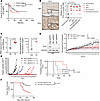 Figure 7
Plk1 knockdown inhibits growth of Bap1-deficient sarcomas. (A) High PLK1 RNA expression correlates with poor prognosis in UPS (TCGA). Survival differences were analyzed using Kaplan-Meier curves and compared using the log-rank test. (B) IHC of a human sarcoma tissue array shows that PLK1 protein expression is significantly increased in UPS, MFS, fibrosarcoma, and dermatofibrosarcoma. Scale bars: 50 μm. Quantification was performed on independent tissue cores, and statistical significance was assessed using a 2-tailed Student’s t test. (C) RNA sequencing and quantitative reverse transcription PCR confirm Plk1 upregulation in PB sarcomas. Statistical comparisons were performed using a 2-tailed Student’s t test. (D) Western blot confirms PLK1 knockdown; cell proliferation assays (CELLCYTE) demonstrate reduced growth following Plk1 knockdown. Statistical comparisons were performed using a 2-tailed Student’s t test. (E) Plk1 knockdown inhibits tumor growth and prolongs survival in a syngeneic mouse model. Survival was analyzed using Kaplan-Meier curves with significance assessed by the log-rank test. (F) CRISPR/Cas9–mediated mutation of Plk1 reduces the penetrance of Trp53/Bap1 loss–driven sarcomagenesis. Survival was analyzed using Kaplan-Meier curves with significance assessed by the log-rank test.
Total RNA sequencing and reverse transcription PCR confirmed that Plk1 was significantly upregulated in PB sarcomas compared with normal muscle (Figure 7C). To investigate the oncogenic role of PLK1, we stably transduced Cas9-expressing PB sarcoma cell lines with lentiviruses expressing either a nontargeting control sgRNA (neg sgRNA) or sgRNAs targeting Plk1. We confirmed reduced PLK1 expression via Western blot (Figure 7D). In vitro proliferation assays showed that Plk1 knockdown significantly inhibited PB sarcoma cell growth (Figure 7D). To assess the effect of Plk1 knockdown in vivo, we injected Rosa26loxP-Cas9/loxP-Cas9 mice intramuscularly with PB sarcoma cells stably expressing neg sgRNA (n = 3), Plk1 sgRNA1 (n = 4), or Plk1 sgRNA2 (n = 3). Plk1 knockdown significantly suppressed tumor growth (Figure 7E). Notably, 2 of 4 mice injected with Plk1 sgRNA1–transduced cells did not develop tumors over a 72-day period. Moreover, Plk1 knockdown significantly prolonged mouse survival (Figure 7E). Next, we examined whether Plk1 upregulation is required for Trp53/Bap1 loss–driven sarcomagenesis. We generated a plasmid expressing 3 sgRNAs targeting Trp53, Bap1 (sgRNA4), and Plk1 (sgRNA1) and delivered it via IVE into the gastrocnemius muscles of Rosa26loxP-Cas9/loxP-Cas9 mice (n = 14). For comparison, a plasmid expressing sgRNAs targeting Trp53 and Bap1 alone was delivered into a separate cohort of mice (n = 24). Additional CRISPR/Cas9–mediated mutation of Plk1 significantly reduced the penetrance of Trp53/Bap1 loss–driven sarcomagenesis (Figure 7F). Collectively, these findings demonstrate that PLK1 is essential for the development and maintenance of Bap1-deficient sarcomas and identify PLK1 as a promising therapeutic target.
Pharmaceutical inhibition of PLK1 as a targeted therapy for Bap1-deficient sarcomas. To evaluate the therapeutic potential of PLK1 inhibition in Bap1-deficient sarcomas, we tested the sensitivity of PB mouse sarcoma cells to volasertib, a selective PLK1 inhibitor (60). In vitro assays demonstrated that PB sarcoma cells were sensitive to volasertib in a dose-dependent manner (Figure 8A). To investigate volasertib’s impact on tumor-infiltrating immune cells, we used Rosa26loxP-Cas9/loxP-Cas9 mice injected intramuscularly with PB sarcoma cells. Treatment began when tumors reached ≥100 mm3, with mice randomized to receive volasertib or vehicle (10 mg/kg, 5 d/wk for 1 week) (Figure 8B). Flow cytometry performed on day 4 after treatment revealed significantly increased frequencies of proliferating (Ki-67+) CD8+ T cells in the volasertib group; no difference in CD11b+ cell percentages among CD45+ cells; significantly lower percentages of PMN-MDSCs and TAMs but higher percentages of M-MDSCs in the volasertib group; and higher expression of PD-L1 on PMN-MDSCs and TAMs in volasertib-treated tumors (Figure 8B and Supplemental Figure 12). These findings indicate that volasertib suppresses tumor growth and modulates the tumor immune microenvironment, enhancing CD8+ T cell proliferation and altering myeloid cell composition.
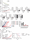 Figure 8
Pharmacologic inhibition of PLK1 suppresses Bap1-deficient sarcomas. (A) Volasertib inhibits PB sarcoma cell proliferation in vitro in a dose-dependent manner. (B) In Rosa26loxP-Cas9/loxP-Cas9 mice, volasertib increases Ki-67+ CD8+ T cells, reduces TAMs, and alters MDSC populations. PD-L1+ PMN-MDSCs and TAMs are elevated after treatment. Statistical comparisons were performed using a 2-tailed Student’s t test. (C) In an autochthonous UPS model, volasertib induces tumor regression in 4 of 6 mice, while niraparib shows no effect. Left: Tumor growth curves. Right: Waterfall plot of tumor size changes after 2-week treatment. (D) Therapeutic efficacy of PLK1 inhibition combined with immune checkpoint blockade in a syngeneic PB sarcoma model. C57BL/6J mice bearing intramuscularly transplanted PB sarcomas were treated with vehicle control (IgG), volasertib alone, anti–PD-1 alone, or the combination. Volasertib was administered by oral gavage (10 mg/kg, 5 days per week), and anti–PD-1 antibody was delivered via intraperitoneal injection (100 μg per dose, days 1, 3, and 5). Combination treatment resulted in greater suppression of tumor growth compared with either monotherapy. Tumor growth curves were compared using 1-way ANOVA followed by appropriate post hoc tests. Kaplan-Meier analysis shows that both monotherapies improve survival relative to control, whereas combination therapy provides the most significant survival benefit. For all experiments, n denotes biologically independent samples or mice as indicated. Data are presented as mean ± SEM unless otherwise specified.
Next, we tested volasertib in spontaneous tumors initiated by IVE delivery of Trp53 and Bap1 sgRNAs into the gastrocnemius muscle of Rosa26loxP-Cas9/loxP-Cas9 mice (Figure 3A). When tumors reached 50–100 mm3 (≥100 days after injection), mice were treated with either 50 mg/kg niraparib (n = 3) or 10 mg/kg volasertib (n = 6) for 2 weeks. Niraparib showed no therapeutic effect, consistent with transplantation model results. In contrast, volasertib induced tumor regression in 4 of 6 mice, with one tumor remaining stable and one showing slow growth (Figure 8C, waterfall plot). Overall, these results demonstrate that pharmaceutical inhibition of PLK1 via volasertib is a promising targeted therapy for Bap1-loss sarcomas, with both direct antitumor effects and beneficial modulation of the tumor immune microenvironment.
Finally, we evaluated whether PLK1 inhibition could enhance the therapeutic efficacy of immune checkpoint blockade in vivo using immunocompetent C57BL/6J mice bearing intramuscularly transplanted PB sarcoma cells. When tumors reached approximately 100 mm3 (day 14 after injection), mice were randomized into 4 treatment groups: vehicle plus control IgG (n = 5), volasertib alone (oral gavage, 10 mg/kg per dose, 5 days per week for up to 2 weeks; n = 6), anti–PD-1 alone (intraperitoneal injection, 100 μg per dose on days 1, 3, and 5; n = 10), and the combination of volasertib and anti–PD-1 using the same dosing regimens (n = 7) (Figure 8D). Relative to the control group, both volasertib and anti–PD-1 monotherapy significantly delayed tumor growth. Notably, the combination treatment resulted in greater tumor growth inhibition compared with either monotherapy. At the end of the treatment period (day 13), tumors were not detectable in mice treated with volasertib; however, tumor regrowth was observed in 4 mice following cessation of volasertib monotherapy. In contrast, mice receiving the combination therapy exhibited more durable tumor control, with tumors remaining undetectable for up to 51 days after treatment initiation. Consistent with these observations, both volasertib and anti–PD-1 monotherapy significantly improved survival compared with control treatment, whereas combination therapy produced the greatest survival benefit (Figure 8D). Collectively, these findings indicate that PLK1 inhibition can enhance the antitumor activity of immune checkpoint blockade in this PB sarcoma model.


